Locations:

Objective clinical and radiographic findings are central to optimal outcomes
By Justin R. Abbatemarco, MD; Chen Yan, MD; Amy Kunchok, MBBS; and Alexander Rae-Grant, MD
Advertisement
Cleveland Clinic is a non-profit academic medical center. Advertising on our site helps support our mission. We do not endorse non-Cleveland Clinic products or services. Policy
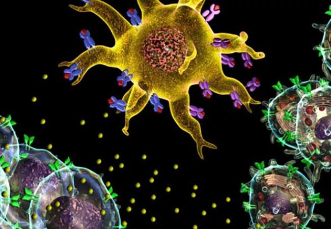
The spectrum and understanding of antibody-mediated autoimmune encephalitis (AE) — an umbrella term for a group of noninfectious, inflammatory central nervous system diseases — have expanded dramatically over the past few years. These disorders typically involve subacute, progressive neuropsychiatric symptoms with associated cognitive dysfunction, movement disorders and autoimmune seizures.
We recently published a review of AE in the Cleveland Clinic Journal of Medicine (2021;88:459-471) focusing on AE syndromes involving cell surface and synaptic proteins. The latter portion of that review — which provides a practical approach to diagnosis and management, including common pitfalls associated with nonspecific antibody findings — is excerpted below. For the complete open-access review, with full references, click here.
When AE is highly suspected, initial testing should include an antibody panel. Both serum and cerebrospinal fluid (CSF) should be tested for antibodies since CSF is more sensitive and specific for certain antibodies, such as NMDA receptor IgG, GAD65 IgG and GFAP IgG, whereas serum is more sensitive for other antibodies, such as LGI1 IgG and Caspr2 IgG.
Antibody panels are preferred over specific antibody tests, given the often overlapping clinical syndromes and the possibility of multiple positive antibodies.
The differential diagnosis for encephalitis is very broad and includes infections, toxic-metabolic encephalopathy, mitochondrial disorders, nutritional deficiencies, vascular disorders, malignancy and demyelinating disorders. Clinicians should be particularly concerned about ruling out an infectious process in view of the immunotherapies used to treat AE. Infections to consider include herpes simplex virus encephalitis, human herpesvirus 6, human immunodeficiency virus, fungal infection (e.g., cryptococcal), mycobacterial infection, Whipple disease and neurosyphilis.
Advertisement
In general, viral infections usually cause a more profound CSF pleocytosis (white blood cell count of 50-100/μL). Bacterial or mycobacterial infections can have a lower CSF glucose concentration, whereas AE usually is characterized by normal glucose levels.
Overall, careful examination usually reveals subtle neurologic deficits that should prompt further evaluation for AE. Diagnostic red flags include newly occurring epileptic seizures, movement disorders and neurocognitive symptoms, especially in the setting of MRI or CSF abnormalities.
It should be mentioned that the prevalence of primary psychiatric disorders is much higher than the prevalence of AE. For example, the overall prevalence of schizophrenia, with incidence peaking in young adults, is estimated to be 2.7 to 8.3 per 1,000 (Psychiatr Clin North Am. 2007;30:323-338), whereas the overall prevalence of AE is 13.7 per 100,000 (Ann Neurol. 2018;83:166-177). Thus, in a young patient with new mood disorder, a primary psychiatric diagnosis remains more likely than AE.
While patients with a preexisting psychiatric disorder can develop a concomitant autoimmune condition, it should also be mentioned that a specific isolated psychiatric AE phenotype has not emerged in the literature despite extensive investigations.
A comprehensive evaluation for suspected AE includes a range of laboratory studies and imaging:
Advertisement
The development of broad antibody panels has led to some unintended consequences, particularly as these panels may include disorders of the central nervous system and the peripheral nervous system. In addition, the specificity and sensitivity can vary for the different autoantibodies and the association with AE. Therefore, interpretation of autoantibody testing should be combined with a comprehensive clinical evaluation as outlined above.
Caution is particularly needed when interpreting tests for autoantibodies that have low specificity for AE. For example, anti-voltage-gated potassium channel (VGKC) antibodies were initially detected in peripheral nerve hyperexcitability disorders such as neuromyotonia and Morvan syndrome. Further laboratory evaluation has elucidated that LGI1 and Caspr2 are the usual targets of these complex antibodies and not the VGKC itself. VGKC antibodies alone are not specific for AE and can be seen in 5% of healthy controls. In VGKC-positive patients without LGI1 and Caspr2 antibodies, additional testing is not usually warranted, as there is no clear evidence that VGKC titers are indicative of an autoimmune disorder.
Hashimoto encephalopathy, also known as steroid-responsive encephalopathy associated with autoimmune thyroiditis, has been historically linked to elevated serum levels of thyroid peroxidase antibodies. In the era of more neuronal-specific antibodies, thyroid peroxidase antibodies have been found to have limited diagnostic value. An extensive evaluation to exclude other causes of AE should be pursued in cases with elevated thyroid peroxidase antibody titers, given the unclear clinical significance of these antibodies in neurologic disorders.
Advertisement
Current treatment guidelines for AE are based on a combination of expert opinion, case series and case reports; evidence from high-quality multicenter randomized trials is lacking. But existing evidence suggests that early initiation of therapy and prompt escalation to second-line immunotherapy may lead to improved clinical outcomes.
Still, unanswered questions include the time frame associated with a response to the first-line immunotherapy and the optimal duration of sustained immunotherapy. A comprehensive evaluation for malignancy is also vital, as early detection and treatment are important for improved patient outcomes.
An important caveat for all clinicians is that although response to corticosteroid therapy is a typical feature of AE, it does not confirm the diagnosis of AE. Disorders such as lymphoma and vasogenic edema associated with a brain tumor can also respond dramatically to steroids. Additionally, some patients with AE do not respond to corticosteroids but require prolonged treatment with other immunotherapies.
Initiation and escalation of treatment depend on the pretest probability of AE along with the clinical severity. If there is a high pretest probability of a known syndrome, waiting for the results of antibody testing should not delay treatment. For example, a case of refractory autoimmune-mediated status epilepticus in the intensive care unit requires prompt and aggressive treatment. Caution should be used when the diagnosis is less certain.
First-line treatments. First-line treatment for AE involves corticosteroids combined with either IVIg or plasmapheresis:
Advertisement
Second-line treatments. Second-line treatments can be given as monotherapy or in combination for refractory disease activity 1 to 2 weeks from completion of first-line treatment:
Maintenance treatments:
There are no validated biological markers to assess treatment response in AE. Although some studies have suggested that early reduction in titers can correlate with better clinical outcomes, antibodies can remain positive even in patients with good outcomes. Further, the change in antibody titers does not consistently correlate with risk of relapse. Therefore, serum or CSF antibody titers in isolation are not reliable for monitoring treatment response in AE.
Imaging, when abnormal, can be repeated to look for improvements over time. Unfortunately, even after appropriate treatment is initiated for AE, brain MRI may show irreversible changes such as generalized or focal atrophy on follow-up.
Monitoring of the treatment response is therefore primarily based on the clinical examination. We urge the use of objective measures to determine the true efficacy of a given treatment. For example, clinicians can use the Scale for the Assessment and Rating of Ataxia (SARA), the Symbol Digit Modalities Test or the Montreal Cognitive Assessment (MoCA) for reliable scores to measure the success of a trial and the utility of long-term treatments. Formal neuropsychological testing is also a valuable tool for documenting the extent of cognitive damage and evaluating immunotherapy response.
Paraneoplastic AE can occur in association with an underlying malignancy. In particular, small-cell lung cancer, non-small-cell lung cancer and neuroblastoma trigger the production of paraneoplastic autoantibodies. The neurologic sequelae can occur prior to detection of the cancer, allowing for early discovery and oncologic treatment.
Though treating the cancer is the main goal in these patients, they may still require short- or long-term immunotherapy for the paraneoplastic disorder. Immunotherapies that may have dual benefit to treat the cancer and autoimmune disorder, such as cyclophosphamide, can be considered.
All treatment decisions must be made in coordination with the treating oncologist. Additionally, patients may have permanent neurologic deficits with relatively little hope for recovery.
The type and frequency of screening for malignancy depend on the specific antibody syndrome. If a patient is diagnosed with an autoantibody commonly associated with paraneoplastic syndromes, frequent monitoring is recommended. In those cases, we typically order surveillance testing every 6 to 12 months for at least 3 to 5 years. If the patient has an antibody with a low likelihood of an underlying neoplasm, we consider reevaluating once a year for 3 years after the index diagnosis.
Based on the 2011 European Federation of the Neurological Societies guidelines, we will use either whole-body FDG-PET or CT of the chest, abdomen and pelvis as a screening measure for certain AE patients, with clinical consideration of the known cancer associations, cancer risk factors or family history of cancer.
In particular, the nature of the antibody determines the risk and type of an underlying malignancy and therefore the investigation. For example, anti-Yo autoantibody has a greater than 90% association with breast or ovarian malignancy, so an extensive evaluation should be pursued to identify the underlying neoplasm.
In addition, sex-specific tests such as pelvic ultrasonography, mammography and testicular ultrasonography should be considered if the initial evaluation is unrevealing. Further, all patients should complete routine age- and sex-appropriate screening measures including screening for breast, colorectal, cervical and prostate cancer, along with lung cancer when applicable, based on U.S. Preventive Services Task Force recommendations.
Our understanding of AE has expanded dramatically over the past few years. Familiarity with different AE syndromes will ensure prompt diagnosis and treatment. In cases that are less clear, a sound diagnostic approach anchored on objective clinical or radiographic findings is important for optimizing outcomes. Clinicians need to stay abreast of developments in this field as the breadth of immune-mediated disorders of the nervous system continues to evolve.
For the complete and fully referenced version of this review, see Cleve Clin J Med. 2021;88:459-471.
Dr. Abbatemarco and Dr. Kunchok are staff neurologists and Dr. Rae-Grant is consultant staff with Cleveland Clinic’s Mellen Center for Multiple Sclerosis Treatment and Research. Dr. Yan is a staff neurologist with Cleveland Clinic’s Center for General Neurology.
Advertisement

Aim is for use with clinician oversight to make screening safer and more efficient

Rapid innovation is shaping the deep brain stimulation landscape

Study shows short-term behavioral training can yield objective and subjective gains

How we’re efficiently educating patients and care partners about treatment goals, logistics, risks and benefits

An expert’s take on evolving challenges, treatments and responsibilities through early adulthood

Comorbidities and medical complexity underlie far more deaths than SUDEP does

Novel Cleveland Clinic project is fueled by a $1 million NIH grant

Tool helps patients understand when to ask for help