Locations:





AI-enhanced mammography improves cancer detection and may support more personalized screening strategies
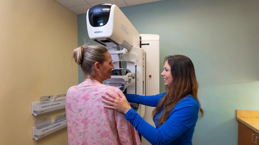
Image content: This image is available to view online.
View image online (https://assets.clevelandclinic.org/transform/16f9ecbd-8382-46bf-8cc2-bbd7c8212d9f/IRH-Vero-radiology-Mammography_9375_SG)
Patient mammography appointment
For decades, breast cancer screening has largely been guided by age. But advances in artificial intelligence (AI), growing recognition of individual risk factors and ongoing debate over screening recommendations are accelerating a shift toward a more personalized approach to early detection.
Advertisement
Cleveland Clinic is a non-profit academic medical center. Advertising on our site helps support our mission. We do not endorse non-Cleveland Clinic products or services. Policy
While professional societies continue to differ primarily on screening intervals and, in some cases, the age at which average-risk women should begin screening, researchers are exploring how AI-enhanced imaging can improve cancer detection and eventually help tailor screening recommendations to an individual's risk profile.
At Cleveland Clinic Indian River Hospital, that future is already beginning to take shape. The hospital’s breast imaging center continually invests in advanced breast imaging technology to improve screening capabilities. Most recently this includes AI-enhanced digital breast tomosynthesis (3D mammography) to support radiologists during image interpretation. Rather than replacing physician expertise, deep learning software functions as a clinical decision support tool that highlights suspicious findings, quantitatively assesses breast density, and provides case and lesion scores.
"AI is a useful tool for radiologists, but it's not in any way a substitution for a radiologist," says Cimmie Shahan, MD, one of four breast radiologists at Indian River Hospital in Vero Beach, Florida. “I think it will have an important role to play as we continue to transition from a one-size-fits-all screening model to a more risk-based approach along with shared decision-making.”
Growing evidence indicates deep learning systems, which are used to recognize complex imaging patterns and generate decision-support insights, can improve breast cancer detection without substantially increasing recall or false-positive rates.
Advertisement
Sweden's MASAI trial found a 29% increase in cancer detection (based on 2025 data) when AI-supported screening was compared with standard double reading by radiologists, with only nonsignificant increases in recall and false-positive rates.
Because U.S. screening practices typically rely on a single interpreting radiologist rather than double reading, these findings may not translate directly. However, the ASSURE study – the largest real-world evaluation of AI-assisted breast cancer screening in the United States – also demonstrated improved performance, reporting a 21.6% increase in cancer detection when AI was added to 3D mammography.
For Dr. Shahan, reinforcement of clinical judgment is among the technology's greatest advantages.
"Many times if there's an area that I think may be concerning on a mammogram and AI agrees, I am reassured that a callback for additional imaging workup is warranted," she says. "At other times, AI may identify subtle abnormalities that prompt a closer review."
Evidence also suggests women with dense breasts benefit from AI-assisted mammography, including findings from the ASSURE study. Although AI cannot overcome the masking effect of fibroglandular tissue, it can improve detection of subtle imaging abnormalities such as architectural distortion and faint microcalcifications that might otherwise be overlooked.
The technology also shows promise as a second reader for general radiologists who interpret breast imaging less frequently, potentially helping to standardize interpretations across practice settings. In addition, AI may eventually help prioritize more suspicious screening examinations in high-volume practices like Indian River Hospital Vero Radiology, although Dr. Shahan says additional evidence is needed before it can be adopted routinely as a triage tool.
Advertisement
She also points out an important limitation. Most current AI software does not compare screening examinations with prior mammograms, and it may flag longstanding benign findings or postoperative changes that an experienced breast radiologist can recognize as stable. However, some newer AI software platforms are incorporating prior mammograms into their algorithms.
Beyond improving image interpretation, AI may eventually help redefine how breast cancer screening recommendations are made.
Current screening recommendations are largely based on age, with additional consideration given to clinical risk factors. Researchers are now developing AI algorithms capable of analyzing mammographic tissue patterns to estimate a woman's future breast cancer risk.
A Swedish retrospective study published in Radiology earlier this year demonstrated that three AI systems identified imaging features associated with approximately 20% of breast cancers up to six years before clinical diagnosis.
Last summer the U.S. Food and Drug Administration granted De Novo authorization for an AI platform that predicts a woman's 5-year risk of developing breast cancer from existing mammograms.
"I believe AI will eventually help us determine the most appropriate screening interval and the most beneficial supplemental imaging modalities based on a patient's level of risk," predicts Dr. Shahan.
She envisions a transition over the next decade from predominantly age-based recommendations toward individualized screening strategies that integrate clinical history, breast density and AI-derived risk prediction. Until then, clinical risk assessment remains the cornerstone of personalized screening.
Advertisement
The movement toward personalized screening comes as professional organizations continue to debate the optimal screening schedule for average-risk women.
Dr. Shahan notes that most organizations agree that annual screening beginning at age 40 saves the greatest number of lives. Where they differ is in how to balance that benefit against potential harms, including patient anxiety associated with callbacks, overdiagnosis and healthcare resource utilization.
The American College of Physicians' updated 2026 guidance continues to recommend biennial mammography for average-risk women ages 50 to 74 and shared decision-making for women ages 40 to 49. The U.S. Preventive Services Task Force also recommends biennial screening but beginning at age 40 and continuing through age 74.
By comparison, the National Comprehensive Cancer Network, American Society of Breast Surgeons, American College of Radiology, and Society of Breast Imaging recommend annual mammography beginning at age 40. Dr. Shahan encourages clinicians to educate patients about both the benefits and potential harms of screening while incorporating individual risk and patient preferences into shared decision-making.
Early risk assessment is especially important because women identified as high risk may begin screening as early as age 25. Cleveland Clinic Breast Radiology currently uses the Tyrer-Cuzick model, although other validated tools, including the Gail model, are also appropriate for initial risk assessment.
Risk assessment also guides decisions regarding supplemental screening.
Advertisement
Current recommendations from the American Cancer Society and the American College of Radiology support annual mammography combined with annual contrast-enhanced breast MRI for women at high risk, ideally alternating every six months for continuous surveillance.
“For patients unable to undergo MRI with contrast, whole breast ultrasound or contrast-enhanced mammography are alternatives,” adds Dr. Shahan.
Intermediate-risk patients, often with dense breasts and additional risk factors, may be considered for supplemental MRI, which outperforms ultrasound, though guidelines are less definitive for this group. For average-risk women with dense breasts, supplemental ultrasound is less strongly recommended due to high false positive rates, though it remains an option for informed patients.
"The breast imaging community is still determining the best course of action for supplemental screening for women who are of average or intermediate risk for breast cancer," says Dr. Shahan, noting that more research is needed.
Rather than viewing AI as simply another imaging tool, Dr. Shahan believes its greatest potential lies in helping clinicians tailor screening recommendations.
"As AI becomes more sophisticated, we'll be able to better identify who is at highest risk, who may benefit from supplemental imaging, and ultimately provide more personalized care," she says.
While that vision will require additional validation and longer-term clinical evidence, Dr. Shahan says the direction is clear. The future of breast cancer screening will increasingly combine clinical risk assessment, physician expertise and AI-supported imaging to deliver more personalized recommendations aimed at detecting cancers as early as possible.
Advertisement
Cleveland Clinic in Florida explores extended reality for multimodality image guidance

The American Society of Clinical Oncology (ASCO) recently released updated guidelines

Cleveland Clinic's transplant team in Florida is using circulatory-death donor hearts to expand transplant opportunities for patients.
Repair of tiny lymphatic vessels offers big relief for patients with lymphedema after cancer surgery
The Center for Neurological Restoration offers a structured, multidisciplinary approach to conditions that often require longitudinal management and escalation beyond first-line therapies
Multidisciplinary expertise provides advanced evaluation, second opinions and personalized treatment planning for patients with challenging cerebrovascular disorders
Study highlights strong predictive value of circulating tumor DNA testing

Researchers with Cleveland Clinic in Florida highlight need for a national registry for gastrointestinal stromal tumors
 Search