Locations:

Review of our recent experience shows it’s still a safe option
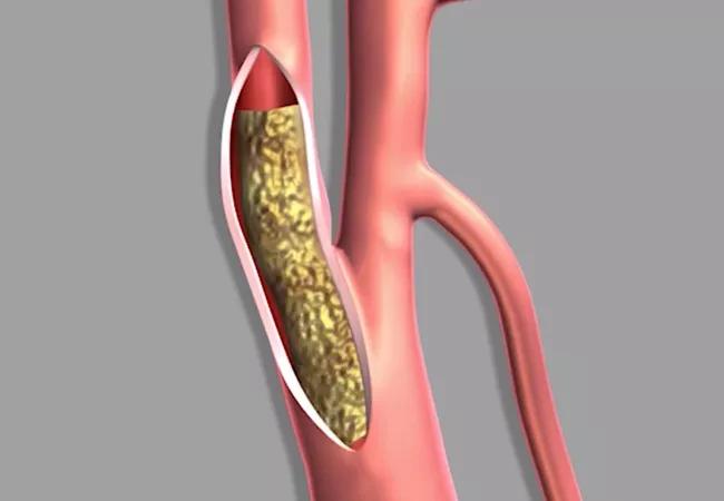
Image content: This image is available to view online.
View image online (https://assets.clevelandclinic.org/transform/a3f21b96-f512-4645-80d9-99489631d6ed/20-HVI-1998312_carotid-endarterectomy_650x450_jpg)
20-HVI-1998312_carotid-endarterectomy_650x450
Carotid endarterectomy (CEA) remains a safe treatment option for severe carotid artery stenosis even in patients at high surgical risk, concludes a large retrospective study from Cleveland Clinic published online by the Journal of Vascular Surgery.
Advertisement
Cleveland Clinic is a non-profit academic medical center. Advertising on our site helps support our mission. We do not endorse non-Cleveland Clinic products or services. Policy
While CEA has been the gold standard for carotid disease since the 1950s, it has been joined more recently by minimally invasive transfemoral carotid artery stenting and transcarotid artery stenting, termed “TCAR” for transcarotid artery revascularization.
Patients with anatomic or physiologic conditions that put them at high surgical risk are often referred for these endovascular alternatives as the least invasive — and presumably safest — procedures. But is there evidence to support this preference? Researchers led by Cleveland Clinic vascular surgeon Francis Caputo, MD, decided to find out by evaluating their institution’s 10-year experience with high-risk patients undergoing traditional CEA.
“We focused on high-risk patients who were considered for CEA or transfemoral carotid artery stenting but who ultimately underwent CEA,” says Dr. Caputo, the study’s senior author. “We found that patients with one or more high-risk factors can undergo CEA and end up with stroke rates comparable to those with transcarotid artery revascularization.”
The analysis focused on 1,347 consecutive patients who underwent CEA at Cleveland Clinic between 2008 and 2018. Among this sample, 1,152 met inclusion criteria for the analysis. These patients were separated into high-risk and standard-risk categories based on whether they had any of various physiologic and anatomic risk factors.
Physiologic risk factors were an ejection fraction < 30%, severe pulmonary disease or an abnormal stress test. Anatomic risk factors were prior head/neck radiation, prior ipsilateral neck surgery, contralateral nerve palsy, redo CEA, prior ipsilateral stenting, contralateral occlusion, contralateral CEA, nasotracheal intubation or a requirement for digastric muscle division.
Advertisement
Initial analysis revealed 450 patients who had one or more high-risk factors. When propensity score matching was used to pair these patients with those without high-risk factors, adequate matches were found for 424 high-risk patients (94%), of whom 173 met at least one physiologic high-risk criterion and 293 met at least one anatomic high-risk criterion.
When the high-risk and standard-risk groups were compared on the primary outcome — a composite of stroke, myocardial infarction or death at 30 days — there were no significant differences in either the composite endpoint or any of its components. Notably, the stroke rate was 1.9% in standard-risk patients versus 1.4% in high-risk patients.
Moreover, results on the primary outcome and its components were comparable between patients with one high-risk factor and those with multiple high-risk factors.
“Since the establishment of high-risk criteria, there have been studies to both support and question the safety of CEA in high-risk patients,” notes Sean Lyden, MD, Chair of Vascular Surgery at Cleveland Clinic and a co-author of the study. “Our findings support the safety of high-risk CEA in centers of excellence.”
The authors observe that rates of stroke, myocardial infarction and 30-day mortality among high-risk CEA patients in their study were comparable to rates among standard-risk patients who received TCAR in a recent review of the Vascular Quality Initiative database. They note that while their study showed high-risk patients to be significantly more likely than standard-risk patients to have a cranial nerve injury, most such injuries were temporary.
Advertisement
“From this analysis,” says Dr. Caputo, “we conclude that CEA remains an effective and safe surgical solution for high-risk patients. Whereas the emergence of transcarotid artery revascularization will reduce demand for transfemoral carotid artery stenting, CEA continues to be a viable option for high-risk patients who fall outside the indications for transcarotid artery revascularization.”
Advertisement
Advertisement

A snapshot of key stats from one of the world's busiest centers

‘Sac flow’ is more precise and will ease unfounded patient concerns, experts argue

Join us in New York Dec. 4-5 for evidence-based instruction with real-world examples

First-ever transcarotid artery revascularization trial with no strokes or device-related deaths

Consensus statement outlines the team, infrastructure and experience needed to deliver TTVI safely and effectively

Innovative approach to living-tissue AVR achieves low reintervention rates, excellent long-term survival

Diagnosis and treatment of malnutrition and cachexia are key to improving cardiac outcomes

Symptom burden at presentation is a potent predictor of long-term survival, large analysis shows