Locations:





Dotatate PET/CT scan and molecular testing informed treatment trajectory
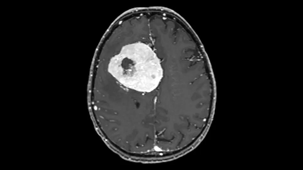
Image content: This image is available to view online.
View image online (https://assets.clevelandclinic.org/transform/d27d3738-c54d-4270-9366-e6d2bc514063/brain-scan-of-brain-large-meningioma)
Large meningiolma
A 49-year-old male patient traveled from his home in Peru to Cleveland Clinic Cancer Institute after being diagnosed with secondary brain cancer. Within days, he was able to receive surgery to remove the large tumor.
Advertisement
Cleveland Clinic is a non-profit academic medical center. Advertising on our site helps support our mission. We do not endorse non-Cleveland Clinic products or services. Policy
At the age of five, the patient was diagnosed with grade 4 medulloblastoma. His oncology team in Peru prescribed the standard of care at the time, which was an intense regimen of surgery, craniospinal radiation and chemotherapy. Given his young age, he was likely to suffer from many late effects, including neurocognitive dysfunction, decreased growth, and hormone dysregulation. The treatment saved his life but put him at risk of secondary cancer.
His parents never told him of his cancer diagnosis or treated him differently. As an active family, they involved him in sports at an early age. “I believe that the fact that he got right back into being active with a healthy lifestyle and athletics impacted him dramatically,” says Erin Murphy, MD, a radiation oncologist with Cleveland Clinic Cancer Institute. “Nowadays we have so many resources like speech therapy, physical therapy occupational therapy and IEPs but at the time he was diagnosed there weren’t as robust resources. Taking advantage of these resources really maximizes young patients’ recovery and how they interact in their everyday lives.”
Forty-four years later, the patient went to the local ER in his hometown due to a puzzling neurological episode. The hospital didn’t have imaging, so he got himself to a medical center in Lima, Peru, where a brain MRI revealed a six-centimeter meningioma that was putting considerable pressure on his brain.
The patient was determined to find the best treatment possible and after much
research, decided to travel to Cleveland Clinic for a consult. After receiving a complete exam and meeting with Dr. Murphy and neurosurgeon Matthew Grabowski, MD, to discuss treatment, he was scheduled for surgery at Cleveland Clinic within days. Dr. Grabowski performed a near total resection of the growth to remove as much of the malignancy as possible while avoiding nearby blood vessels. The 12-hour surgery was a success, and the patient was discharged from the hospital earlier than expected.
Advertisement
The surgical pathology showed the growth was consistent with meningioma and Neuro-oncologist Alejandro Torres-Trejo, MD, arranged for a sample to be sent for molecular testing. The patient wanted to return to Peru to be with his young children while awaiting the molecular test results.
Based on this desire, the care team worked with him to coordinate care with his medical oncology providers in Peru. The plan was for him to get an MRI locally at home and have it sent to the Cleveland Clinic team for review. At that point, the care team would regroup and determine if further intervention was warranted.
The MRI indicated potential progression, which was concerning since it’s unusual for a grade 1 meningioma to grow that quickly. This was an early indication that radiation therapy was appropriate. However, the patient contracted a wound infection and needed to stay close to home until that resolved.
In the meantime, the molecular imaging results had returned. The tests showed markers for a grade 2 or atypical meningioma. “There’s still no consensus in the medical community of how to treat grade 2 meningiomas, but what we now know is that what cells look like under the microscope doesn’t tell the whole story. This is a good example where molecular information can really help guide treatment,” says Dr. Murphy. Once the wound infection healed, the patient was able to return to Cleveland to start radiation therapy.
The patient’s care team continued its extensive workup, including a dotatate PET/CT scan, which is very specific imaging for this type of cancer. This sophisticated imaging technique helped personalize the radiation dosage based on the specific patient’s situation.
Advertisement
The scan revealed several areas of concern outside the resection cavity. Due to the areas of concern in the scan as well as the molecular testing, the plan was for the patient to undergo 6.5 weeks of comprehensive irradiation to treat the residual disease and the entire resection cavity. Since there was residual disease around the tumor site, the patient received both a standard dose of radiation to the resection cavity as well as a simultaneous integrated boost of radiation to the areas of residual tumor cells.
The patient was motivated to stay active throughout his treatment. He retained a positive attitude throughout the many weeks of daily radiation. His wife accompanied him to Cleveland and joined him on daily walks. Two weeks after completing radiation therapy, he was well enough to go on a much-anticipated European vacation with his family.
He continues to be monitored closely by his medical oncologist back home. His Cleveland care team also arranged for distance health visits to minimize the need to travel back to the states. He’ll receive MRIs near his home and continue to have virtual visits with Cleveland Clinic. If any of the small remaining lesions grow, he would be a candidate for a less-invasive Gamma Knife® radiosurgery.
Dr. Murphy shared several lessons learned from this complex case.
“Dotatate PET scans are very helpful for finding residual disease and checking for any other possible meningiomas. They can help guide us, giving us the option to deliver an additional boost of radiation to areas of residual disease.”
Advertisement
“I always tell patients the better you get good nutrition, the more active you are
and the better you stay hydrated, the better you’ll be able to get through radiation therapy. This patient was walking miles every day, eating well and enjoying life. He barely had any side effects. He was very positive and very driven. It made such a difference.”
“Anyone who gets a new diagnosis understandably wants to know all the facts immediately, but molecular testing and advanced imaging make workups take longer, and things can change along the way as you learn more. It’s important to set expectations for our patients that we continue to undertake fact finding so we know all that we can about them and their tumor so that we know how best to treat them.”
Advertisement
Advertisement

New program will help patients improve fitness and manage weight after cancer treatment
Cleveland Clinic hematologist highlights new guidelines and treatment advances in immune thrombocytopenia
NIMBLE replaces weekly tumor board delays with same-day multidisciplinary recommendations
Cleveland Clinic study provides validation for the test, which combines AI and pathological data to predict outcomes in postoperative patients
Advanced imaging technology catches metastases much earlier, allowing treatment with fewer side effects

Reduced radiation for lymph node areas reduces side effects
Large Cleveland Clinic analysis may help resolve a longstanding question over optimal fractionation
This single-port option offers advantages for appropriate patients
 Search