Locations:





Multicenter study offers guidance on when to consider extended monitoring
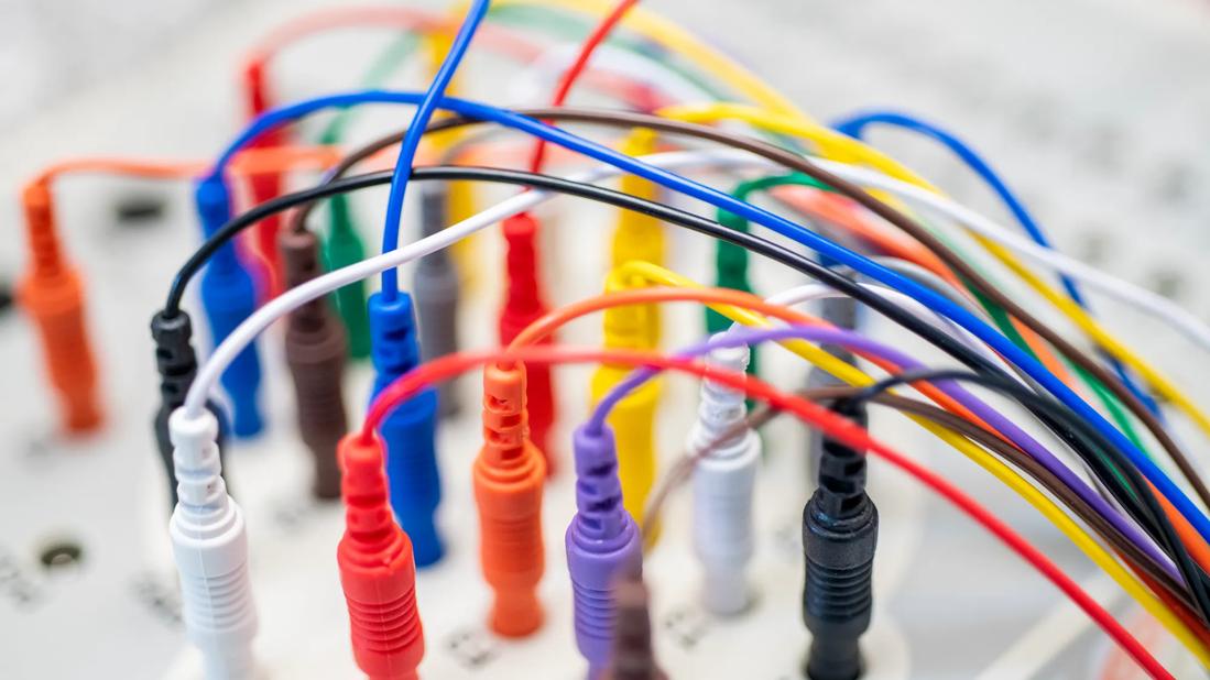
Image content: This image is available to view online.
View image online (https://assets.clevelandclinic.org/transform/3626796d-7985-44b2-9103-77e22fee9f31/NEU_2865231_06-02-22_028_LDJ)
multicolored EEG wires plugged into an electrode junction box
In the acute post-stroke setting, extending EEG monitoring to at least 12 hours significantly improves detection of intermittent epileptiform patterns and provides superior predictive value for post-stroke epilepsy compared with standard 60-minute recordings.
Advertisement
Cleveland Clinic is a non-profit academic medical center. Advertising on our site helps support our mission. We do not endorse non-Cleveland Clinic products or services. Policy
That’s the conclusion of a recent retrospective study published in Annals of Neurology (2026;100[2]:400-415) based on patients from Cleveland Clinic and Hôpital Universitaire de Bruxelles in Belgium.
“The question of which patients with acute ischemic stroke warrant prolonged EEG monitoring has been a bit of a conundrum, especially when it comes to its value in informing long-term outcomes like post-stroke epilepsy” says co-principal investigator Vineet Punia, MD, of Cleveland Clinic’s Epilepsy Center. “Our findings suggest that selective use of continuous EEG can better identify high-risk stroke survivors who lack clinical seizures but may still develop chronic epilepsy. This type of approach should allow for more accurate patient counseling and can help prioritize limited neurophysiological resources toward patients who stand to gain the most from extended monitoring.”
Stroke remains the foremost cause of epilepsy in older populations, accounting for more than half of new-onset cases after age 65. While clinical acute symptomatic seizures are a recognized risk factor, they occur in only a small portion of patients who eventually develop post-stroke epilepsy. “There is a real need for alternative biomarkers to identify at-risk survivors early in their recovery,” Dr. Punia notes.
He and other researchers from the current study demonstrated in a recent investigation (Ann Neurol. 2025;98:814-825) that EEG results within seven days after stroke are strong independent predictors of post-stroke epilepsy, especially in patients without acute symptomatic seizures. That investigation led them to propose the SeLECT-EEG prognostic score for post-stroke epilepsy development.
Advertisement
Despite that insight, the optimal duration of EEG monitoring has been unclear. Short EEGs lasting 20 to 60 minutes are common and broadly feasible, but they often miss transient or intermittent abnormalities. Continuous EEG (cEEG) — i.e., lasting 12 hours or more — has been shown to be more sensitive in detecting these abnormalities. Prior to the current study, the specific prognostic benefit of moving from a short EEG to cEEG for assessing long-term seizure risk had not been rigorously quantified in a post-stroke cohort.
The new retrospective study analyzed 283 adults with acute ischemic stroke from two centers from the SeLECT-EEG investigation — Cleveland Clinic and Hôpital Universitaire de Bruxelles. To be included, patients had to undergo cEEG within seven days of their stroke and have no history of prior epilepsy or acute symptomatic seizures.
The researchers used a robust “within-recording” comparison design. For each patient, they simulated a 60-minute short EEG by taking the first hour of the patient’s cEEG and compared those findings against the full continuous record, which lasted at least 12 hours. This ensured that differences in findings were strictly due to monitoring duration rather than variations in electrode placement or clinical state. All EEGs were reviewed using standardized terminology by board-certified neurophysiologists who were blinded to patients’ long-term outcomes.
The study revealed that cEEG uncovers significantly more pathologic activity than a one-hour study. Specifically, continuous monitoring increased the detection of several key indicators, including:
Advertisement
Beyond detection of abnormalities, the findings had implications for long-term prognosis. Over median follow-up of 41 months, 14.5% of the cohort developed post-stroke epilepsy. The strongest predictors of future epilepsy were lateralized periodic discharges and electrographic seizures, both of which were detected at least twice as often on cEEG.
The researchers also evaluated monitoring duration in terms of its effect on the SeLECT-EEG score, which incorporates stroke severity, etiology and EEG findings to predict the risk and timing of post-stroke epilepsy. When this score was calculated using cEEG data, it achieved a significantly higher C-index (0.68) than when using short EEG data (0.63), indicating greater predictive strength. Additionally, the emergence of any epileptiform activity after the first hour of monitoring was associated with a 28% risk of developing epilepsy within five years, compared with a risk of only 11% in patients in whom such activity was never found.
The study authors contend that their findings offer practical guidance for when to extend EEG monitoring beyond the initial hour. They found that patients with a SeLECT2.0 score of 4 or higher, indicating higher baseline clinical risk, derived the most prognostic benefit from continuous monitoring.
In their study report, the researchers propose a matrix for classifying patients into four categories to guide decisions around the use of cEEG:
Advertisement
Notably, no patient with a completely normal cEEG developed post-stroke epilepsy during the follow-up period. “This suggests that a normal continuous EEG might eventually serve to largely rule out long-term epilepsy risk, though this requires confirmation in broader populations,” Dr. Punia notes.
The authors conclude that continuous monitoring is not just a more sensitive version of routine EEG but also provides unique prognostic data that can help reclassify future seizure risk in stroke survivors. They urge stroke units to move toward a risk-guided, phenotype-based approach to cEEG in this setting to ensure the most appropriate use of this specialized monitoring.
They add that while current management guidelines do not recommend routine prophylactic antiseizure medications in this setting, accurately identifying high-risk patients is the first step toward future trials of preventive therapies and more structured follow-up care for post-stroke epilepsy.
Advertisement
Advertisement
Where the field is moving and what Cleveland Clinic has in the works
Collaboration with AI startup promises to reshape neurocritical care monitoring at scale

Highest response rates seen with stress-lowering techniques like yoga, meditation and mindfulness

Free portal helps researchers classify and share data using the IC-CoDE framework
Large study shows rural patients are less apt to be discharged to inpatient rehab, hampering outcomes

Inferior clinical outcomes continue into mechanical thrombectomy era, large analysis finds
Quantitative imaging adds diagnostic value beyond 3T MRI in nearly half of patients
Data-driven segmentation approach shows promise for seizure characterization with utility for clinical decision making
 Search