Locations:

Consider 36-hour standard, with certain risk factors prompting longer or shorter windows
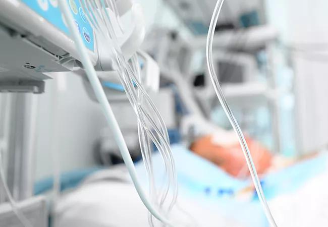
Image content: This image is available to view online.
View image online (https://assets.clevelandclinic.org/transform/c1d2320f-cc53-4f09-a3ec-21c7fb148d65/21-NEU-2612260-critical-care_650x450_jpg)
Medications in the ICU. Medicinal lines next to patient bed
It may be time to extend the standard time window for continuous EEG (cEEG) monitoring among critically ill patients, suggests a large retrospective Cleveland Clinic study published in Epilepsia Open.
Advertisement
Cleveland Clinic is a non-profit academic medical center. Advertising on our site helps support our mission. We do not endorse non-Cleveland Clinic products or services. Policy
The study found that among critically ill patients undergoing cEEG monitoring, seizure detection increases linearly for the first 36 hours, with detection after that time more likely to occur in patients who are taking antiseizure medications or who have altered mental status, periodic epileptiform discharges or an acute brain insult.
“Based on the linear trend to 36 hours that we found for seizure detection in critically ill patients, we recommend extending the current cEEG monitoring standard of 24 hours,” says the study’s senior author Stephen Hantus, MD, Director of Continuous EEG and Epilepsy Consult Service at Cleveland Clinic. “And with this large study cohort, we were able to identify risk factors to justify longer — or shorter — monitoring durations.”
Nonconvulsive or subclinical seizures can be detected in up to about 20% of critically ill patients undergoing cEEG monitoring. While most such seizures can be detected within 24 hours of monitoring, others are delayed and therefore easily missed. Current recommendations are to monitor for up to 24 hours for most patients, and for at least 48 hours in patients who are comatose or have a history of seizures. This guidance is based on risk factors identified by other investigations of highly selected patient cohorts.
The current study was designed to identify any additional risk factors for delayed seizure detection by cEEG in a large and more varied patient population.
The study cohort consisted of hospitalized adults who underwent cEEG at Cleveland Clinic during 2016. Of 2,402 patients, 316 (13.2%) had at least one nonclinical seizure. The mean age of the entire cohort was 59.4 ± 17.4 years (49.6% female), and the most common primary etiologies of presentation were toxic/metabolic/infectious encephalopathy, brain bleeds and ischemic strokes. The most common indication for monitoring was an observed seizure-like episode.
Advertisement
Timing of first nonclinical seizure detection by cEEG occurred at the following rates:
Cumulative seizure detection increased linearly for the first 36 hours of monitoring, after which the odds of seizure detection increased by 46% for each additional day of monitoring.
The following were identified as risk factors for delayed seizure detection (i.e., after 48 hours):
Dr. Hantus notes that a patient’s clinical presentation provides some of the most important indicators of optimal duration of cEEG monitoring. Patients who present with an acute brain insult, especially mixed bleeds, are at high risk of delayed seizure detection and need longer monitoring. Likewise, anyone with coma, stupor or lethargy is likelier than patients with awake mentation to need extended monitoring.
The study report outlines the following general guidelines for duration of cEEG monitoring.
Standard monitoring duration should be 36 hours. This is an increase from current published recommendations of up to 24 hours of cEEG for patients who are not comatose and without a seizure history. As 36 hours is the optimal duration among the entire study cohort regardless of risk factors, it is most relevant for patients for whom little clinical history information is available.
Advertisement
For certain patients, up to 24 hours of continuous monitoring is adequate. For awake patients without interictal epileptiform discharges, less than 24 hours monitoring should suffice. Also, although patients who have a witnessed seizure-like event are at high risk for having seizures during monitoring, these are usually detected within 24 hours.
Multiple situations warrant 48 hours of cEEG monitoring. These include:
“Timely seizure detection with cEEG may reduce medical and neurological complications, and even in-hospital mortality,” concludes Dr. Hantus. “Findings from this study can help clinicians make decisions about optimal cEEG monitoring duration with increased confidence.”
Advertisement
Advertisement

Findings may have implications for understanding the disorders’ pathophysiology

Routine capture of standardized neuroperformance data may expand and refine investigations

Nearly one-fifth of such cases fulfill 2024 McDonald criteria based on biomarkers

Adequate dosing may improve outcomes in well-selected patients, large Cleveland Clinic series suggests

Reimagining the outpatient neurological visit with routine capture of neuroperformance data

Free portal helps researchers classify and share data using the IC-CoDE framework

Large study shows rural patients are less apt to be discharged to inpatient rehab, hampering outcomes

Updates on this fast-evolving therapeutic landscape from a leading trialist