Locations:

A pulmonary pathologist explains how to tell the difference
In this video, Sanjay Mukhopadhyay, MD, staff pulmonary pathologist in the Department of Anatomic Pathology, presents an interesting case of Nonspecific Interstitial Pneumonia lung disease. Dr. Mukhopadhyay highlights the differences in Usual Interstitial Pneumonia (UIP) and Nonspecific Interstitial Pneumonia (NSIP), and explains what a pathologist should look for when determining the patient’s diagnosis.
Advertisement
Cleveland Clinic is a non-profit academic medical center. Advertising on our site helps support our mission. We do not endorse non-Cleveland Clinic products or services. Policy
Video content: This video is available to watch online.
View video online (https://www.youtube.com/embed/-PXyberbf4w?feature=oembed)
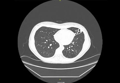
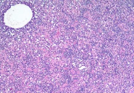
Dr. Mukhopadhyay shows a case of a patient in his 40s with interstitial lung disease and a CT scan showing bilateral reticular opacities with some traction bronchiectasis. He explains that typically, the differential diagnosis for this patient would include both UIP and NSIP after radiology, and pathology would be the determining factor to distinguish between the two. He notes in the biopsy that there appears to be no significant variability in the appearance of the lung, which is a key characteristic of UIP (For another discussion of UIP, watch Carol Farver, MD, director of Pulmonary Pathology, explain the characteristics and pathologic findings). Dr. Mukhopadhyay emphasizes the lack of the characteristic patchwork pattern in the biopsy shown as an important clue.
At higher magnification, the abnormal interstitium is a mix of chronic inflammatory cells as well as collagen in the background. This appearance is different from smoking-related interstitial fibrosis in which the alveolar septa are expanded by ropy hyalinized collagen alone without inflammatory cells. Collagen is a hallmark of NSIP. The uniform distribution of the interstitial fibrosis as well as the presence of collagen and chronic inflammation is characteristic of NSIP.
This video is part of the Pathology Insights series, featuring important cases, methods and practices personally presented by staff pathologists. The short videos break down information about interesting pathology cases to better inform doctors, laboratory staff, patients or anyone interested in the field of pathology.
Advertisement
Advertisement

Screen patients seeking care for chlamydia, gonorrhea
Lingulectomy removes infection when antibiotics fail
Researchers have developed immunoprofiles for an emerging disease with a mortality rate as high as 27%

Findings from one of the first published case series
Don't discount this crucial step
EBUS-TBNA found safe and effective
How to spot the rare infection
Not all lung nodules are malignant