Locations:





Cleveland Clinic in Florida explores extended reality for multimodality image guidance
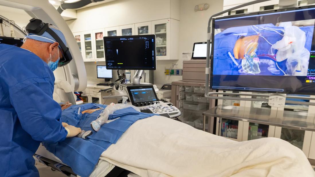
Image content: This image is available to view online.
View image online (https://assets.clevelandclinic.org/transform/ab7ec2d7-facb-4578-b324-cd8441f36204/WH-Mediview-XR90_Procedure_1747_CROPPED2_SG-2)
MediView’s XR90™ holographic surgical navigation system
Interventional radiologists rely on sophisticated imaging technology to perform precise, image-guided procedures with minimal disruption to the body. They must translate two-dimensional (2D) images into a three-dimensional (3D) understanding of the patient's anatomy and then use that information to guide instruments precisely to the intended location. That can be challenging when multiple imaging modalities are needed to access deep structures.
Advertisement
Cleveland Clinic is a non-profit academic medical center. Advertising on our site helps support our mission. We do not endorse non-Cleveland Clinic products or services. Policy
New extended reality (XR) technology at Cleveland Clinic Weston Hospital offers another approach. The hospital is the first in Florida to acquire and deploy the MediView XR90™ surgical navigation and intelligence platform, which combines preprocedural CT imaging with real-time ultrasound in one 3D holographic view via a see-through XR headset.
As an early adopter of the technology, Cleveland Clinic Weston Hospital is evaluating how XR navigation can complement conventional image-guided techniques, initially for biopsies and potentially for other needle-based procedures.
“It’s a tool that's forecasting where medicine is going in the coming years,” says Kevin Stadtlander, MD, an interventional radiologist and Site Director of Cleveland Clinic’s Interventional Radiology Residency Program at Weston Hospital.
Image-guided procedures traditionally require physicians to move between information on imaging monitors and the patient while maintaining a mental picture of how the target relates to surrounding structures.
The XR90 system approaches that challenge by combining information from different imaging modalities into a patient-specific 3D representation.
Preprocedural CT imaging provides detailed anatomic information that is fused with live ultrasound during the procedure and spatially registered to the patient's anatomy. This is projected as a 3D holographic overlay into the physician's field of view through a heads-up display.
The system uses skin-marked fiducial locations, specialized physical registration markers and electromagnetic tracking to establish the spatial relationship between the digital images and the patient, with sub-centimeter targeting accuracy.
Advertisement
“The system overlays information onto the real world, allowing me to see both the patient and digital data at the same time,” says Dr. Stadtlander, the first at Weston Hospital to use the technology.
The hospital's first cases with the XR90 system are helping physicians evaluate where multimodality XR guidance may offer the greatest benefit.
In one case, the patient had a small liver lesion approximately 10 cm beneath the skin and located between two branches of the hepatic vein. This location presented a challenge for percutaneous biopsy using only one imaging modality.
Instead, Dr. Stadtlander used the system to combine the patient's preprocedural CT images with real-time ultrasound. The 3D visualization showed the lesion and planned needle trajectory in relation to surrounding anatomy, while ultrasound provided live information during needle advancement.
The value of the technology, reports Dr. Stadtlander, lies in how it changes the way information is presented during a procedure. Instead of relying solely on separate 2D images, the physician can view the target and relevant anatomy in a shared 3D environment.
“This is particularly useful when lesions are anatomically challenging or difficult to localize using a single imaging modality,” he says.
The potential advantages of XR navigation extend beyond enhanced visualization and improved precision.
During conventional image-guided procedures, physicians repeatedly shift their gaze between the patient and monitors positioned elsewhere in the procedure room. With the headset, relevant imaging information remains within the physician's field of view.
Advertisement
“Wearing the headset to view all the necessary information directly in front of my eyes reduces neck and back discomfort from constantly looking around for screens,” says Dr. Stadtlander.
The approach also brings preprocedural CT information into the procedure without requiring additional CT imaging during the intervention. That access to detailed anatomic information potentially limits the need for additional exposure to ionizing radiation for patients and members of the clinical team.
Cleveland Clinic Weston Hospital's initial clinical use of the XR90 system will focus on percutaneous biopsies, particularly cases in which multimodal imaging could assist with targeting. Potential future applications may include thermal ablation and other procedures.
The goal, Dr. Stadtlander says, is straightforward: “If we can see better, we can do better.”
By using the platform in both clinical care and research, clinicians will provide feedback that may help refine future applications of the technology and identify where it provides the greatest value. The technology also could have applications beyond individual procedures by enhancing physician education and promoting collaboration among specialists across the Cleveland Clinic system.
“As advanced imaging systems, artificial intelligence and robotics continue to expand the capabilities of image-guided medicine, we remain focused on optimizing patient care,” adds Dr. Stadtlander.
Advertisement
Advertisement
The American Society of Clinical Oncology (ASCO) recently released updated guidelines

Cleveland Clinic's transplant team in Florida is using circulatory-death donor hearts to expand transplant opportunities for patients.
Repair of tiny lymphatic vessels offers big relief for patients with lymphedema after cancer surgery
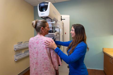
AI-enhanced mammography improves cancer detection and may support more personalized screening strategies
The Center for Neurological Restoration offers a structured, multidisciplinary approach to conditions that often require longitudinal management and escalation beyond first-line therapies
Multidisciplinary expertise provides advanced evaluation, second opinions and personalized treatment planning for patients with challenging cerebrovascular disorders
Study highlights strong predictive value of circulating tumor DNA testing

Researchers with Cleveland Clinic in Florida highlight need for a national registry for gastrointestinal stromal tumors
 Search