Brillouin imaging, corneal cross-linking could aid identification and management of keratoconus and postoperative ectasia

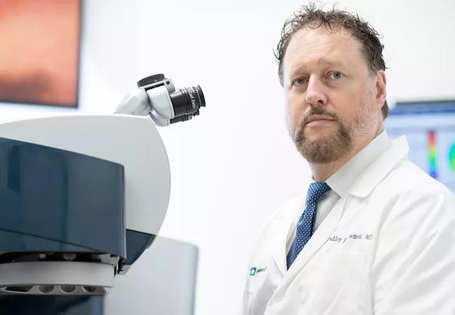
In 2019, internationally known corneal and intraocular refractive surgeon and researcher J. Bradley Randleman, MD, joined the staff of Cleveland Clinic’s Cole Eye Institute.
Advertisement
Cleveland Clinic is a non-profit academic medical center. Advertising on our site helps support our mission. We do not endorse non-Cleveland Clinic products or services. Policy
Dr. Randleman previously held the Hughes Professorship of Ophthalmology at Emory University, and later was Professor of Ophthalmology at the University of Southern California’s Keck School of Medicine and Director of the Cornea & Refractive Surgery Service at USC’s Roski Eye Institute. He is Editor-in-Chief of the Journal of Refractive Surgery and has written four textbooks and more than 150 peer-reviewed publications.
Dr. Randleman’s research focus is the identification and management of corneal ectatic diseases, including keratoconus and postoperative ectasia after LASIK, and the therapeutic potential of corneal cross-linking (CXL). He is the co-principal investigator of a five-year, $2 million National Institutes of Health Research Project Grant (R01) to develop an optical technology that could improve keratoconus diagnosis and treatment.
Daniel F. Martin, MD, predicts that, with the combination of Dr. Randleman, Ocular Biomechanics and Imaging Laboratory Director William J. Dupps, MD, PhD, and Director of Corneal Research Steven Wilson, MD, the institute will become a major center of expertise in corneal research.
Consult QD spoke with Dr. Randleman about his work and plans.
Q: Why did you choose keratoconus as a research focus?
Dr. Randleman: I just found it fascinating. It was a bit of a mystery. You can’t physically see the anatomical changes that patients are having until they’re pretty far down the road, diagnostically. The more I learned about it, the more I realized how little we truly understand keratoconus at the basic physiologic, anatomic level, and even causally, why certain people get keratoconus and others don’t. Why it progresses so far, so fast, in some patients and barely changes in others. It’s tied in nicely with the other side of my clinical practice, which is refractive surgery, because that’s the major thing we’re screening for — any subtle signs of an ectatic cornea.
Advertisement
Q: How has refractive surgery changed with the realization that keratoconus can be an unintended result?
Dr. Randleman: The indications for surgery have shrunk a bit since the 1990s. We don’t treat as high myopia as we initially did. We certainly screen much more diligently than we did in the early period.
Q: What’s the prevalence of keratoconus, both spontaneous and surgery-induced?
Dr. Randleman: For disease-based keratoconus, the best estimate is about one in 2,000, but that’s for fairly late-stage disease. It varies tremendously by region. We did a study of prevalence in pediatric patients in Riyadh, Saudi Arabia, and it was one in 21. A study in the Netherlands found one in 375 in the general population. It’s not unreasonable to think that as many as one in 500 people have some evidence of disease, and maybe as many as one in 200 have some subtle, abnormal findings. For post-refractory surgery prevalence, one in 2,500 to 3,000 is a ballpark figure. Maybe as little as one in 5,000, depending on the surgical center.
Q: You were involved in developing an early method of quantitatively assessing patients’ risk for ectasia after LASIK surgery. How did that come about, and what’s the status now?
Dr. Randleman: When LASIK was introduced in the U.S. in the late 1990s, there were some initial reports of postsurgical ectasia. People wondered whether it was an anomaly or could it happen to anyone, and was it predictable based on risk. Our study was published in 2008 and found that no single factor did particularly well at identifying everyone at risk. It was a combination of variables — abnormal topography was the most significant, but also low residual stromal bed thickness, thinner preoperative corneas, higher-myopia treatments and younger patient age. The good news is that most of the things that were evident at that juncture are screened out ahead of time. The vast majority of refractive surgeons avoid operating on topographies that fell into the suspicious range. There are multiple different imaging technologies available now to assess patients that were not available at the time we developed that weighted risk-assessment approach. Scheimpflug imaging, epithelial thickness mapping, and more recently, optical coherence tomography-based imaging, have given us a dramatically better understanding of corneal anatomy.
Advertisement
Q: What about Brillouin imaging?
Dr. Randleman: Brillouin imaging is fascinating. It’s used in astronomy and a variety of other fields. In ophthalmology, it’s being used to evaluate the cornea, lens, and even the lamina cribrosa. Shining a light on the cornea produces a shift in the light frequency emitted from corneal tissue based on the tissue interaction. If you have a high-enough intensity laser and a sensitive-enough sensor, you can measure those changes and essentially determine the stiffness of the tissue, not just overall but by layer.
Q: For your NIH grant research, you and co-principal investigator, University of Maryland physicist Giuliano Scarcelli, are using Brillouin imaging to better understand corneal biomechanics in keratoconus. Tell us more.
Dr. Randleman: First, we want to look at a wide range of patients in their baseline state — normal patients and those with various stages of keratoconus, from subclinical to advanced — to see what information Brillouin imaging can give us. Our hope is that we can find some biomechanical metrics that identify these eyes as being different than normal in places where our current imaging does not. The second focus of our study is using Brillouin to assess individual refractive procedures that we know weaken the cornea, to determine the amount of weakening, where it happens, and to what extent it differs in various procedures. How different, biomechanically, is LASIK versus PRK versus SMILE [small-incision lenticule extraction]? And finally, with cross-linking, which we know strengthens the cornea in keratoconus, we’ll use Brillouin to see how quantifiable the change in stiffness is before and after cross-linking and where it occurs anatomically.
Advertisement
Q: How would you use that information?
Dr. Randleman: If we’re able to answer those questions, it allows us to start looking at individualizing refractive surgery protocols or cross-linking protocols. If we can tell a patient coming in what their corneal biomechanical profile is, we can better match them with a specific treatment protocol.
Q: How would you do that? Are you hoping the research results in a diagnostic device?
Dr. Randleman: I think the outcome will be a clinically available device. That’s certainly where we’re headed. Ultimately, the hope is that we can find something that will give us a sense of the cornea’s inherent strength, so that we know if we can modify it with refractive surgery or if we need to be thinking about doing a strengthening procedure before the patient has had any real anatomic changes and lost any vision. And, ideally, we’ll even be able to determine how much treatment needs to be done to give an exact outcome. We’re many steps away from that. First we need to characterize what’s happening en masse to the cornea.
Q: Corneal cross-linking, or CXL, is a relatively new approach to strengthen the cornea, approved by the U.S. Food and Drug Administration in 2016 to treat progressive keratoconus and post-LASIK ectasia. Are there still outstanding questions about it?
Dr. Randleman: Certainly one thing we don’t know is how long the effect lasts. The earliest treatments were around 2000, in Dresden, Germany. We’re getting reasonably good 10-year data. We’ll be happier when we have 30-year data. The biggest thing that confounds us right now is trying to predict what type of outcome an individual patient will have. The vast majority of patients are stabilized with CXL, meaning their corneas do not continue to get steeper or thinner, but not everyone is. Who are those outliers? Did they need more treatment? A higher intensity? Longer treatment? Those things we don’t know.
Advertisement
Q: Is that outcome variability due to individual variation in patients, possibly in corneal biomechanical properties? Or to variation in the CXL protocol, such as whether it’s done with the epithelial layer intact or removed?
Dr. Randleman: My first guess is that it’s something different about the individual patient. Any epi-on protocol is doing something to supposedly enhance riboflavin penetration, and that’s poorly studied to date. Even in the 96% of patients who have stabilization with CXL after epithelial-off treatment, some have significant flattening, some have no flattening, some have an induction of astigmatism, and some have a reduction of astigmatism. So all of those are things we would like to understand better and, ideally, to treat individually, which we just don’t have the ability to do right now.
Q: Does CXL have potential uses beyond treating ectasia?
Dr. Randleman: The big one is in infectious keratitis. That’s really exciting, because a single treatment may be as effective as weeks of antibiotics. And it takes out that guesswork of whether it’s gram-positive, gram negative or an early fungal infection. We also don’t have to worry if a particular patient is going to be resistant to one medicine versus another. Once infections become deeper and later-stage, cross-linking is less effective. It probably would be best as a frontline treatment. There have been some other potential CXL uses discussed. Later-stage treatment for patients with corneal edema who are not good surgical candidates —it may have some limited role there. Another is cross-linking for refractive outcomes and/or corneal regularization. Those are quite exciting.
Q: What do you mean by regularization?
Dr. Randleman: For instance, maybe you have a patient who is coming in for cataract evaluation and has some irregular astigmatism. You don’t need to stabilize their cornea, but if you could reduce their irregularity by selectively applying crosslinking, that would be a great minimally invasive procedure that may improve their vision as well. CXL also has been looked at as a less-invasive option for low myopia and low hyperopia. The jury is still way, way out on that. But if we became a bit more refined in those, then people with half a diopter of refractive error who don’t really generally go for surgery may become refractive surgery candidates using this selective CXL treatment. And for people who have a good but not perfect refractive outcome after cataract surgery, CXL may be a noninvasive treatment we could do to fine-tune their vision. Another area that’s in desperate need of improvement is primary keratoprostheses. These devices are a mix of plastic or some type of nonorganic material combined with a corneal graft. One of the big issues is that the corneal rim is subject to melting. Some doctors have cross-linked those. It hasn’t been done very often, but making keratoprostheses more robust would be a real game-changer.
Q: Why did you decide to join Cole Eye Institute’s staff?
Dr. Randleman: It’s a phenomenal place. I had the good fortune of working with Dr. Martin when he was at Emory University School of Medicine. He was on faculty when I started my residency training. He’s always been a friend and mentor. I’ve collaborated with Steve Wilson and B.J. Dupps numerous times on studies, panels and course designs. Dr. Dupps and I are looking at the same thing — corneal biomechanics — from different directions. Dr. Wilson is predominantly looking at wound healing — corneal microstructure, if you will — which is intimately important in what we’re doing with cross-linking, in how we may modify refractive procedures based on what we’re trying to do biomechanically. So these things all fit really well together. There’s really no place in the United States that is this well-funded from a research standpoint and has this many individuals working on refractive surgery, keratoconus and the continuum of corneal biomechanics. I’m very excited to be here.
Advertisement

Motion-tracking Brillouin microscopy pinpoints corneal weakness in the anterior stroma

Registry data highlight visual gains in patients with legal blindness

Prescribing eye drops is complicated by unknown risk of fetotoxicity and lack of clinical evidence

A look at emerging technology shaping retina surgery

A primer on MIGS methods and devices

7 keys to success for comprehensive ophthalmologists

Study is first to show reduction in autoimmune disease with the common diabetes and obesity drugs

Treatment options range from tetracycline injections to fat repositioning and cheek lift