Locations:

Advances in imaging technology could offer new insights for combatting age-related muscle loss
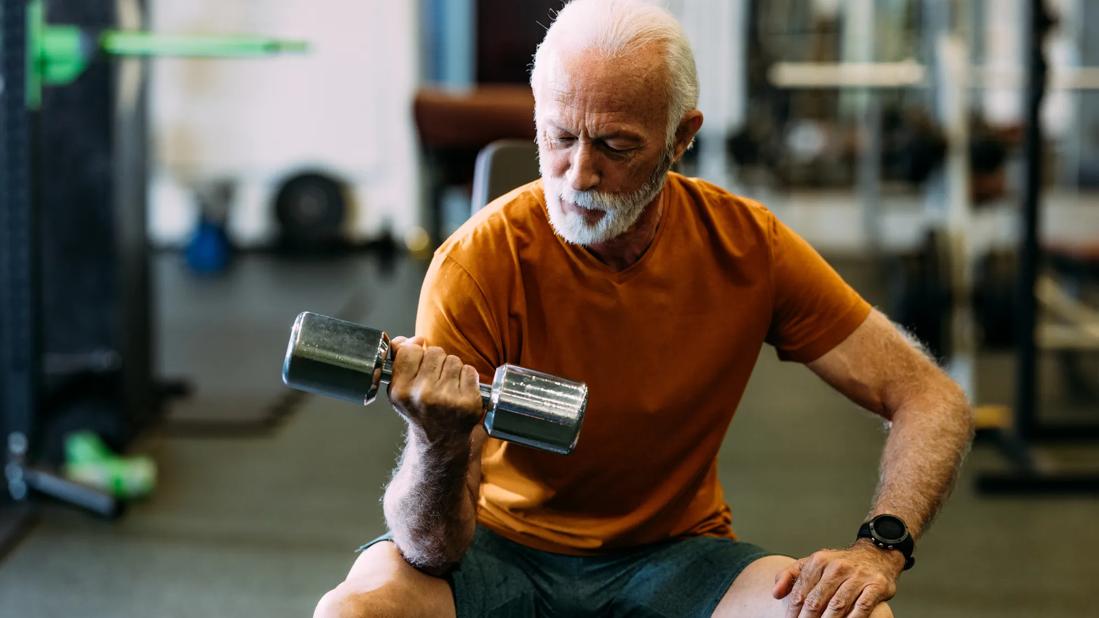
Image content: This image is available to view online.
View image online (https://assets.clevelandclinic.org/transform/afd50e5f-d6d5-43e1-a7a4-b618a0043730/elder-male-lifting-weights-2155616069)
older man lifting weights
By 2050, the global population of people over age 80 will triple, reaching 426 million, according to the World Health Organization. As life expectancy increases, so does the prevalence of age-related conditions, including sarcopenia. Sarcopenia affects mobility, increases the risk of falls, and contributes to frailty and other comorbidities.
Advertisement
Cleveland Clinic is a non-profit academic medical center. Advertising on our site helps support our mission. We do not endorse non-Cleveland Clinic products or services. Policy
Understanding sarcopenia’s underlying mechanisms is crucial to developing interventions that preserve strength and function in an ageing population.
Recent advancements in MRI technology have enhanced the ability to assess muscle composition, providing increasingly detailed insights into sarcopenia’s progression and its relationship to overall health. These imaging techniques enable researchers to evaluate both muscle quantity and quality, supporting the development of tailored treatment approaches.
At the forefront of this research is Alister Hart, MA, MD, FRCS (Orth), a consultant trauma and orthopaedic surgeon at Cleveland Clinic London, working in collaboration with University College London and the Royal National Orthopaedic Hospital. His multidisciplinary team of surgeons, biomedical engineers, biotechnologists and radiologists explores how exercise influences musculoskeletal health.
One of the team’s key studies, published in BMJ Open Sport & Exercise Medicine, examined the impact of marathon training on knee health. Approximately 70 first-time marathon runners underwent MRI scans before and after a four-month standardised training programme. Two senior musculoskeletal radiologists assessed the runners’ internal knee structures using validated scoring systems.
“Our study found that pre-existing subchondral bone marrow oedema showed reversibility following the training and marathon, challenging long-held assumptions about the impact of running on joint health,” Prof Hart says.
Advertisement
A similar study, published in the Orthopaedic Journal of Sports Medicine, assessed hip health in people who had MRI scans of both hips 16 weeks before and two weeks after running their first marathon. Results showed that labral tears, bone marrow oedema and other abnormalities of the tendons and ligaments visible on MRI did not impact running performance.
“MRIs after the marathon showed very few changes from the MRIs before the marathon, indicating that running one marathon is not associated with aggravating hip injuries or increasing hip pain,” Prof Hart says.
Another study by Prof Hart’s team, published in Skeletal Radiology, reported no adverse effects of marathon training and running on the lower lumbar spine.
The team also used MRI to study the impact of cycling on muscle health. One study, published in BMC Musculoskeletal Disorders, compared middle-aged men (average age 49) who cycled recreationally (training for an average of 15 years and cycling more than 7,000 km in the previous year) with those who were physically inactive. Research results showed that the cyclists had lower levels of fat in their gluteal muscles and had larger muscles than men who were inactive.
“Our previous research suggested that fat infiltration in the gluteus maximus could be a valuable marker of muscle health, good mobility and healthy hips,” Prof Hart says. “Strong muscles are known to help protect joints.”
To advance their study of MRI and muscle health, Prof Hart’s team has developed artificial intelligence (AI) tools to automatically segment core and lateral hip muscles on MRI images. These tools enable accurate measurement of cross-sectional area and fat fraction, offering a more precise evaluation of muscle health, specifically of the lumbar spine muscles, including the psoas, iliacus, quadratus lumborum, erector spinae and multifidus.
Advertisement
The team continues to refine imaging protocols to improve the sensitivity and specificity of MRI in detecting early changes in muscle composition. They also are investigating novel biomarkers that could provide further insight into sarcopenia’s progression.
“Our future research will focus on integrating these imaging techniques into routine clinical assessments, facilitating early detection and intervention strategies that could improve patient outcomes,” Prof Hart says. “By combining advanced imaging with clinical practice, our work aims to develop more effective approaches to managing sarcopenia and preserving muscle function in older adults.”
Advertisement
Advertisement

As genetic insights refine diagnosis, research abounds on current and emerging therapies

Genetic testing at Cleveland Clinic provided patient with an updated diagnosis

Quantitative imaging adds diagnostic value beyond 3T MRI in nearly half of patients

The new application that could augment cancer detection, improve efficiency

Two studies from Cleveland Clinic may help advance the technology toward broader clinical use

Overcoming barriers to implementing clinical trials

Multimodal evaluations reveal more anatomic details to inform treatment

A closer look at the impact on procedures and patient outcomes