Locations:

A look at emerging technology shaping retina surgery
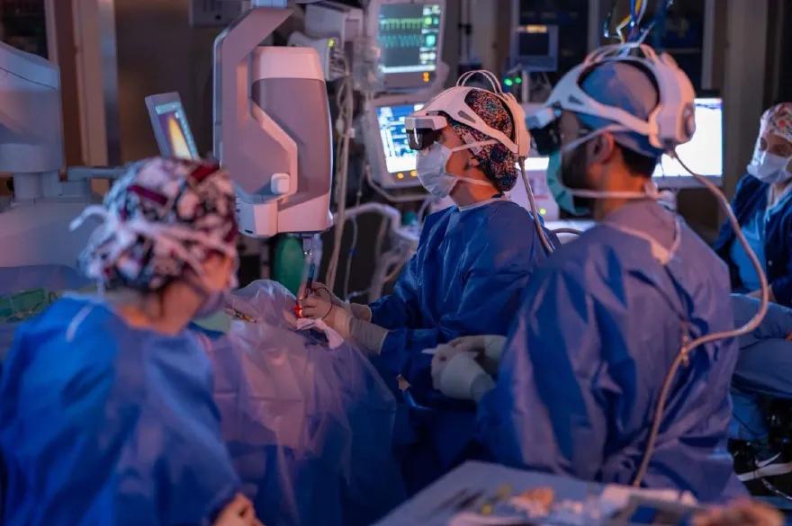
Image content: This image is available to view online.
View image online (https://assets.clevelandclinic.org/transform/cde8b2bc-b7b6-4e55-97cd-443fa15c54aa/heads-up-display-retina-surgery)
Using heads-up display during retina surgery
Advertisement
Cleveland Clinic is a non-profit academic medical center. Advertising on our site helps support our mission. We do not endorse non-Cleveland Clinic products or services. Policy
In a retina clinic, advanced imaging is essential. It’s critical for accurately diagnosing and managing patients with neovascular age-related macular degeneration, a persistent macular hole or proliferative diabetic retinopathy, for example. We use tools like optical coherence tomography (OCT) angiography to identify choroidal neovascular membrane, diabetic macular edema or retinal vein occlusion.
We rely so heavily on images to “see” patients that I can’t imagine functioning in a retina clinic without OCT. However, using these imaging tools intraoperatively is not standard for retina surgery.
Why is that? Should that change?
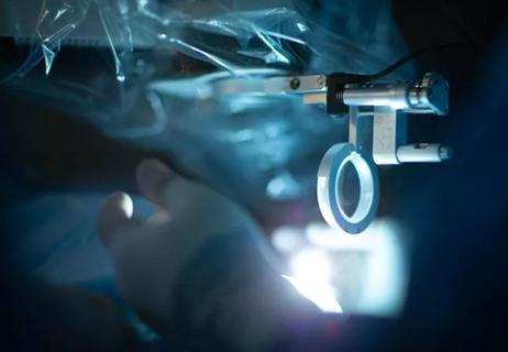
Intraoperative OCT (iOCT) is a valuable adjuvant to ophthalmic surgery. It increases our understanding of disease pathology and can provide real-time feedback to retina surgeons.
For example, when peeling an epiretinal membrane, real-time OCT can reveal traction. When injecting tissue plasminogen activator for submacular hemorrhage, OCT can help us ensure that the cannula is accurately placed, under the retina but not under the subretinal pigment epithelial space. When creating a scleral window for uveal effusion syndrome, OCT can help us determine exact tissue depth.
Intraoperative OCT also can provide information on subtle changes in the retina in response to surgical maneuvers. It can help guide procedures when necessary or help us know when further action is unnecessary.
The prospective PIONEER and DISCOVER studies, led by Cleveland Clinic Cole Eye Institute’s Justis P. Ehlers, MD, showed the feasibility and utility of iOCT in anterior and posterior segment surgery. Out of more than 500 patients in the PIONEER study, successful scans were obtained in 98% of cases, pausing surgery a median of 4.9 minutes. Out of more than 800 patients in the DISCOVER study, successful scans were obtained in 98% of cases, leading to altered decision-making during the procedure in 29% of cases.
Advertisement
For example:
While iOCT offers many benefits, there are some drawbacks:
Vitreoretinal visualization with a standard operating microscope has been relatively stagnant in its evolution. Now advancing surgical visualization are 3D heads-up displays. They provide a digital stereoscopic view of the surgical field on a high-definition monitor, freeing the surgeon from the confines of microscope eyepieces and potentially improving ergonomics and efficiency.
Advertisement
There are multiple systems being developed, including Artevo (Zeiss), NGENUITY® (Alcon), SeeLuma™ (Bausch + Lomb) and Beyeonics. The goal of all these systems is to create an integrative theater where surgeons are immersed in the surgery. While looking ahead at the surgical field and performing the procedure, you also can view measurements such as intraocular pressure and vacuum settings. You can keep your eyes in one place but still get lots of information.
Features of these systems vary. One offers improvements in surgical field color, which is especially helpful when doing macular work or peeling membranes. One offers digital binoculars and a design that allows you to view a heads-up monitor without leaning to look around a tower. One offers augmented reality in which you can focus, pan and zoom the digital microscope with your head movements. When you integrate it with iOCT, you can bring the retina into focus and align iOCT images next to it to really see what’s going on.
There have been studies evaluating the use of these innovative systems. In one prospective study that I worked on in 2018, we compared a standard operating microscope with the NGENUITY 3D heads-up system during macular surgery. We found that the heads-up display required lower endoillumination (which is safer for patients), although the standard microscope was better for surgeons’ ease of use (comfort) and peel time (efficiency).
A 2020 study showed that the NGENUITY 3D heads-up system was better for educating ophthalmology trainees, although some individual steps (e.g., endolaser, scleral fixation of IOL, closing) may be more challenging than with a standard display.
Advertisement
While 3D heads-up systems have the potential to change the way we operate, can the technology replace our standard microscopes without compromise? Can they offer something that we don’t already have, especially for our most challenging cases?
Overall, the shortcomings of these systems include:
Regardless of these shortcomings, I think it’s important to continue to use 3D heads-up systems to help them advance. However, limited funding and scarce space in hospitals and surgery centers will continue to be a challenge. Smaller centers can purchase a limited number of systems, and certain systems may not do all things well for all surgeons and all types of procedures.
Advertisement
Three-year results of the DISCOVER study indicated surgeons’ strong preference for visualizing OCT data on a 2D screen (which required looking away from the surgical field) versus viewing OCT data displayed in microscope oculars. That finding highlighted the need for a new integrative digital surgical field.
In 2020, we compared surgeons’ likelihood of viewing iOCT data on a screen versus the surgical field when using a conventional, microscope-integrated system or a digitally enabled heads-up display during vitreoretinal surgery. We found that surgeons using the NGENUITY heads-up display were more likely to view iOCT images while looking at the surgical field. We found similar results in 2024 with the Artevo heads-up display. These results are likely due to the quality of iOCT images in a 3D heads-up system compared to microscope oculars.
In conclusion, there is a lot of potential for innovation in surgical imaging and visualization that can be beneficial for teaching and ergonomics, not to mention better understanding of retina pathology. These systems may be the future, but they need to offer us something that we don’t have currently. There is a great need for more outcomes-based research to show what can be useful and what can’t.
Dr. Talcott is a vitreoretinal surgeon at Cleveland Clinic Cole Eye Institute. This article is based on her presentation at the 2025 Cole Eye Institute Imaging Summit.
Advertisement

How computational tools and personalized biomechanics can improve keratoconus detection, ectasia risk assessment and surgical outcomes

A multitude of subspecialities offer versatility, variety

Prematurity is linked with poorer outcomes of retinal surgery in adulthood

Therapies include steroid implants, immunomodulation and biologics
Corneal imaging and interpretation play a major role

Cole Eye Institute imaging specialists are equal parts technician, artist and diagnostician
30% of references generated by ChatGPT don’t exist, according to one study
Novel use of tPA reduces fibrin response in the eye