Locations:

A primer on MIGS methods and devices
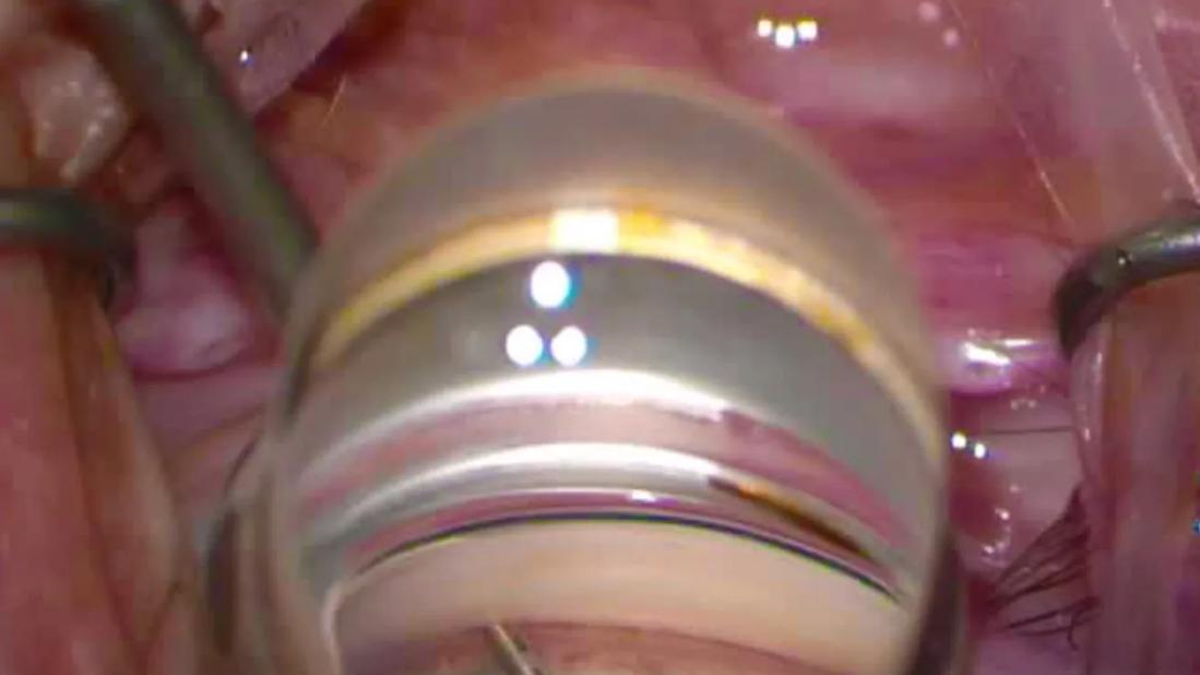
Image content: This image is available to view online.
View image online (https://assets.clevelandclinic.org/transform/f5d3e47a-ee2f-420f-89b8-8b4786a50320/minimally-invasive-glaucoma-surgery)
Microinvasive glaucoma surgery
By Nicole Bajic, MD, and George Markakis, MD
Advertisement
Cleveland Clinic is a non-profit academic medical center. Advertising on our site helps support our mission. We do not endorse non-Cleveland Clinic products or services. Policy
There are various types of microinvasive glaucoma surgery (MIGS) procedures and devices. In order, from the simplest to the most challenging to perform, these include:
This method dilates the canal of Schlemm after creating a small ostomy through the trabecular meshwork. It does not involve further alteration of the trabecular meshwork or the placement of an implant. Each depression of the actuator (maximum 8) creates precise openings in the trabecular meshwork and delivers 7 µL of viscoelastic into the Schlemm’s canal.
The procedure can be done as a stand-alone or in conjunction with cataract surgery. Due to its modest lowering of intraocular pressure (IOP), Streamline is most suitable for mild primary open-angle glaucoma (POAG) and ocular hypertension. This is the easiest MIGS procedure to perform.
There are other forms of canaloplasty (e.g., iTrack™ Advance, Omni®, Via360®) that may offer more robust IOP lowering. However, these other systems generally have a steeper learning curve compared with Streamline.
This effective stand-alone technique can remove a strip of trabecular meshwork four to six clock-hours long. Some patients may develop hypotony.
Because it is a tissue-altering method, it frequently causes hyphema, which can sometimes persist or even spill into the anterior vitreous, especially if a patient is on anticoagulants. The majority of postoperative heme will clear. For this reason, it is generally not necessary to stop anticoagulation therapy, which could increase the patient’s risk of a vascular complication. In these cases, you may find benefit in doing the MIGS before phacoemulsification as well as using a heavier viscoelastic (like Healon®) to tamponade heme.
Advertisement
This technique may be appropriate for ocular hypertension as well as any type and severity of glaucoma: mild, moderate, severe, or open- or narrow-angle (in cases of residual open-angle glaucoma, provided the trabecular meshwork is visible). It may not be the best choice for patients with a high-end intraocular lens (IOL) implant given patient expectations and the potential complications of persistent hyphema, anterior vitreous hemorrhage and hypotony, and how they may affect the long-term performance of IOLs.
This system includes three preloaded 360 µm titanium stents. These stents produce minimal tissue disruption, but generally more modest IOP lowering as well. Since they are rarely complicated by hyphema or hypotony, they can pair well with premium IOLs.
Many consider the iStent as an appropriate first procedure for a surgeon learning to do MIGS. You can go through the main wound, but you need to maintain the ideal position and pressure while inserting the stent. While IOP reduction is more modest with iStent, the procedure typically produces less postoperative reflux, heme and inflammation.
The iStent infinite is the newest generation of iStent, with an infinite number of “clicks” allowing for more opportunities to adjust the stent, if needed.
The procedure involves placing an 8 mm curved stent that has windows for aqueous outflow. It perforates the trabecular meshwork, inserting into and dilating the Schlemm’s canal. This allows aqueous humor to flow unimpeded through the stent, from the anterior chamber to Schlemm’s canal and associated collector channels. As there is a low incidence of complications from hyphema and hypotony, it also can pair well with premium IOLs.
Advertisement
There is a short learning curve when using Hydrus. However, with experience and good anatomy, the procedure can be completed in several minutes. You can practice placement of the Hydrus inserter by using a Sinskey hook. Bury the tip of the inserter into the trabecular meshwork and angle slightly superiorly as you deploy the stent. Do not force it into place. If you feel any resistance, retract the inserter and go further downstream. Use the inserter (turned backhanded and slid within the notch at the end of the stent) or a Sinskey hook to adjust the final position as needed.
These devices are highly effective at lowering IOP by combining two methods to increase aqueous outflow: canaloplasty, which dilates the canal of Schlemm, and incisional goniotomy, which incises the trabecular meshwork. They can be tissue-sparing (e.g., canaloplasty) or tissue-altering (e.g., goniotomy). Up to 12 clock hours can be treated if desired.
Given the options of performing canaloplasty and/or partial or complete goniotomy, these devices are highly customizable and can be titrated to the degree of glaucoma.
The procedures can be performed as a stand-alone or in conjunction with cataract extraction and are effective for both ocular hypertension and most types of glaucoma.
These more advanced techniques are more invasive and tissue-altering. The potential for complications such as hypotony and hyphema is much greater with these procedures and may necessitate additional postoperative care, such as needling. One device (CyPass) was removed from the market after studies demonstrated significant unintended corneal endothelial loss. It is for these reasons that we recommend these procedures be left to glaucoma specialists and other anterior segment surgeons who are more experienced with treating complex glaucoma and its complications.
Advertisement
One way to ease into performing angle surgery is to practice gonioscopy at the end of phacoemulsification so you can get used to viewing, positioning and performing the bimanual technique. Ask the patient if it’s OK to do an extra exam after their cataract surgery so you can learn more about their eye. Then use the Sinskey hook simply to probe the trabecular meshwork.
When you’re ready for your first glaucoma surgery, we suggest you consider starting with Streamline or a goniotomy. After becoming comfortable with these cases, transition to iStent or Hydrus procedures. Use trypan blue to stain the trabecular meshwork in your first few cases. When you are more familiar with angle techniques, pitfalls and patient selection, that can be a good time to explore other procedures.
For 7 keys to success in performing MIGS, see How to Get Started With Microinvasive Glaucoma Surgery (Part 1).
Drs. Bajic and Markakis are comprehensive ophthalmologists at Cleveland Clinic Cole Eye Institute.
Advertisement
Advertisement

Gonioscopy can help determine next steps in reducing IOP

Prescribing eye drops is complicated by unknown risk of fetotoxicity and lack of clinical evidence

7 keys to success for comprehensive ophthalmologists

From medication to laser treatment to surgery

Tissue remnants seem unrelated to clinical outcome

Minimally invasive surgery is effective for uveitic and steroid-induced glaucoma too

The drug does not provide lasting disease control for most patients

New service for patients with diabetes will make annual screening more convenient