Locations:

Key considerations when diagnosing and managing severe hyponatremia
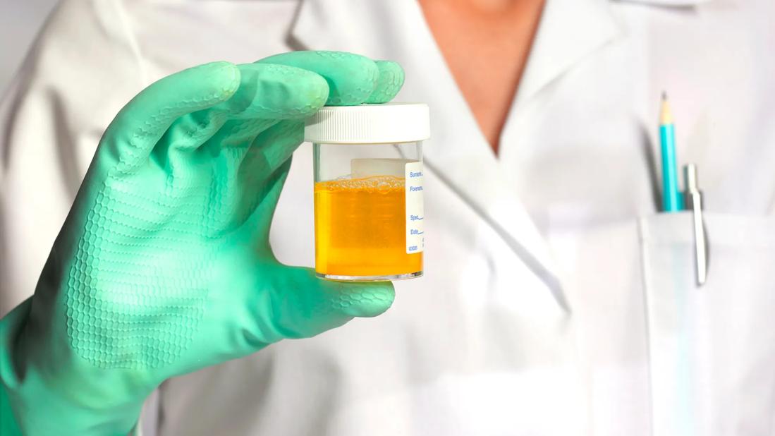
Image content: This image is available to view online.
View image online (https://assets.clevelandclinic.org/transform/617b6e71-5563-405f-b939-4b53f7f8646a/urine-sample-1178770880)
Clinician holding urine sample in gloved hand
Editor's note: This article was originally published in the Cleveland Clinic Journal of Medicine.
Advertisement
Cleveland Clinic is a non-profit academic medical center. Advertising on our site helps support our mission. We do not endorse non-Cleveland Clinic products or services. Policy
By Elias Bassil, MD; Georges N. Nakhoul, MD, Med; Jonathan J. Taliercio, DO, FASN; and Ali Mehdi, MD, MEd, FACP, FASN
A 52-year-old woman was brought to the emergency department by her friend because she was concerned about the woman’s new state of confusion. Her friend provided most of the history. She stated that the patient had not been acting like herself for several days. The patient reported mild nausea and anorexia for the past month, resulting in significant weight loss (estimated at 15 lb), as well as a drastically increased alcohol intake after being laid off from work. The patient was an active smoker and had a 60-pack-year smoking history. She had no other past medical history and was not on any prescription medications.
Vital signs taken in the emergency department were as follows:
No orthostasis was noted. Her weight was 86 kg (189.6 lb), with a body mass index of 32 kg/m2.
Physical examination revealed a disheveled woman in no acute distress. She was alert and oriented only to self. Her mucous membranes were moist. There was no jugular venous distention. Cardiovascular examination was normal, and there was no lower-extremity edema. Lung auscultation revealed bronchial breath sounds over the right lower base. The abdomen was examined and found to be normal. Table 1 lists the results of laboratory testing.

Image content: This image is available to view online.
View image online (https://assets.clevelandclinic.org/transform/4ea01cb4-9cdd-4cae-a57a-faa3febe71b6/Severe-hyponatremia-table1)
Chest radiography revealed a consolidation in her right middle lobe—there was no prior image for comparison. Electrocardiography showed normal sinus rhythm, and computed tomography of the head with contrast showed no acute intracranial abnormalities.
Advertisement
1. What studies would help differentiate the etiology of the patient’s main electrolyte disturbance?
This patient’s clinical picture is consistent with severe hyponatremia, defined as a plasma sodium concentration less than 120 mmol/L or symptoms ascribed to low sodium levels,1 resulting in encephalopathy. Initial evaluation of hyponatremia should include serum osmolality, urine osmolality and urine sodium tests, which ideally should be obtained before initiating therapy.
Serum osmolality
The first step when evaluating hyponatremia is to determine whether the hyponatremia is due to a hypoosmolar state (< 275 mOsm/kg).2 This is referred to as true hyponatremia and is the most common category of hyponatremia. Hyperosmolar hyponatremia (> 295 mOsm/kg), also referred to as translocational hyponatremia, occurs when water shifts out of the intracellular fluid into the extracellular space because of highly osmolar substances in the serum such as glucose and mannitol.
Iso-osmolar hyponatremia (275−295 mOsm/kg) can also be translocational, as seen with moderate hyperglycemia. A more uncommon possibility is pseudohyponatremia, which is seen in hyperproteinemia and hyperlipidemia. Measured hyponatremia in hyperlipidemia and hyperproteinemia is the result of the indirect measurement techniques used in the laboratory. Direct techniques, such as those that employ ion-selective electrodes and blood gas analyzers, would show the true normal sodium concentration.3
Advertisement
Urine osmolality
Urine osmolality is a direct surrogate for the presence of circulating ADH, also referred to as arginine vasopressin. ADH is produced by the supraoptic and paraventricular nuclei in the hypothalamus and stored in the posterior pituitary. Under physiologic conditions, the release of ADH is tightly controlled. Rat studies have demonstrated that ADH is mainly released in response to changes in plasma osmolality and effective circulatory volume.4 Plasma osmolality is tightly controlled, and small changes in osmolality trigger ADH release or suppression. In hypertonic states, osmoreceptors within the hypothalamus signal for ADH release, whereas decreased effective circulatory volume is sensed by various baroreceptors, ultimately leading to hypothalamic ADH secretion.5
Unlike plasma osmolality, a significant change in effective circulatory volume is required to trigger ADH release. However, once the threshold is achieved, the response is much more robust,4 potentially surpassing the response to the osmolality stimulus. States of shock can increase vasopressin levels up to 500-fold.6 When released into the circulation, arginine vasopressin binds to vasopressin V1a receptors, leading to vasoconstriction. Binding to basolateral vasopressin V2 receptors located on the collecting duct of the renal tubule causes the insertion of aquaporin channels into the luminal membrane, which leads to increased free water reabsorption. The net effect is the formation of a concentrated (hyperosmolar) urine and movement of solute-free water from the tubular fluid into the plasma.7
Advertisement
Urine osmolality can range from approximately 50 mOsm/kg to 1,200 mOsm/kg. A urine osmolality higher than 100 mOsm/kg implies ADH-mediated free water reabsorption and would be considered abnormally concentrated urine in a hyponatremic state.8 A urine osmolality less than 100 mOsm/kg denotes a dilute urine and implies the absence of ADH or lack of response to ADH.8 In the context of hyponatremia, the latter is usually seen in low-solute states and in primary polydipsia.
Urine sodium
A random urine sodium test can help evaluate kidney perfusion. A urine sodium level less than 30 mmol/L indicates a sodium-avid state in the face of decreased effective circulatory volume. This occurs in states of true volume depletion or in the hypervolemic states of heart or liver failure or nephrosis. A urine sodium level greater than 30 mmol/L is usually indicative of a sodium-replete state. Diuretic use or impaired renal tubular sodium reabsorption, for example, in Addison disease, can lead to higher urine sodium despite effective hypovolemia. In addition, glucosuria or the presence of urinary anions (as in bicarbonaturia) can also elevate urinary sodium independent of volume status.9
In the context of hyponatremia, a low urine sodium level is usually suggestive of decreased effective circulatory volume. A higher urine sodium level is usually seen in the syndrome of inappropriate ADH secretion (SIADH) and some cases of primary polydipsia.
Random urine potassium
Although measuring urine potassium may be important in guiding management in hyponatremia as it relates to the assessment of the clearance of electrolyte-free water, this test has no diagnostic utility in the initial evaluation of hyponatremia.
Advertisement
Serum ADH measurement
Measuring serum ADH is generally not pursued in the diagnostic workup of hyponatremia. While ADH measurement is offered by some laboratories, ADH is very unstable when isolated from plasma, making measurement and interpretation quite challenging.10 Copeptin, or C-terminal proarginine vasopressin, is generated by enzymatic cleavage of the vasopressin prohormone. It is a more stable alternative and can potentially be measured.10 However, most commercial laboratories do not offer such testing, limiting its utility.
The results of further laboratory testing are listed in Table 2.

Image content: This image is available to view online.
View image online (https://assets.clevelandclinic.org/transform/d92c4296-a917-4019-a6cf-5d62abf80246/Severe-hyponatremia-table2)
2. Based on these results, which of the following is the most likely diagnosis?
SIADH
As reviewed above, increased plasma osmolality and decreased effective circulatory volume are the main stimuli for ADH release. SIADH occurs when ADH is produced in the absence of an osmotic or effective circulatory volume stimulus. This can occur with ectopic ADH release in certain cancers, especially lung cancer, a known paraneoplastic process.11 SIADH can also be seen with many medications including antidepressants, anticonvulsants, and antipsychotics.12 Disorders of the central nervous system, including but not limited to infections, trauma, and metastatic disease, can also result in SIADH.13 It is noteworthy that nausea and pain are potent endogenous stimulants of ADH release, frequently precipitating hyponatremia in the postoperative period, particularly when intravenous fluids are administered.14
The urine studies in our patient are inconsistent with SIADH. The dilute urine, evident by a urine osmolality of less than 100 mOsm/kg, indicates the absence of ADH. This is the appropriate physiologic response to hypotonicity (serum osmolality of 237 mOsm/kg). Contrarily, the inappropriate release of ADH in SIADH (despite the low serum osmolality) would yield a high urine osmolality. Additionally, the urine sodium level would be expected to be higher than 30 mmol/L, appropriately indicating a non-sodium-avid state and suppression of the renin-angiotensin-aldosterone system.15
Hypovolemia
Hypovolemic hyponatremia is caused by ADH secretion triggered by reduced effective circulatory volume. Urine studies are expected to reveal a concentrated urine (urine osmolality > 100 mOsm/kg and usually > 300 mOsm/kg) and a low urine sodium level indicative of the sodium-avid state (except in cases of renal losses due to cerebral salt wasting or Addison disease, as detailed below). The high urine osmolality in these scenarios is reflective of ADH secretion aimed at reestablishing a depleted effective circulatory volume. In our patient, there were no clinical signs of overt volume depletion. In addition, the low urine osmolality would not support this hypothesis.16
Liver cirrhosis
Liver cirrhosis results in a hypervolemic state accompanied by a decreased effective circulatory volume. This results in the maladaptive release of ADH and the resultant hyponatremia commonly seen in persons with cirrhosis.17 Urine studies will show a low urine sodium level (indicative of the sodium-avid state due to low effective circulatory volume) and an elevated urine osmolality (> 100 mOsm/kg) due to ADH secretion. A similar physiologic response occurs in advanced heart failure. Notably, hyponatremia indicates a poor prognosis in heart failure and cirrhosis.18,19 Although the patient had elevated transaminases along with an aspartate transaminase-to-alanine transaminase ratio of 2:1, suggesting alcoholic hepatitis, there were no signs of hypervolemia or any stigmata of liver cirrhosis. Moreover, as in true hypovolemia, elevated urine osmolality along with a very low urine sodium level would be expected.
Cerebral salt wasting
Cerebral salt wasting is a potential cause of hyponatremia in patients with an underlying central nervous system pathology. It is characterized by renal sodium losses leading to hypovolemia.20 ADH release is stimulated by hypovolemia and a decreased effective circulating volume. As such, urine osmolality in these patients is increased. In contrast to other causes of hypovolemia, urine sodium levels in cerebral salt wasting are quite elevated given that the renal sodium leak is the reason for the observed hypovolemia.21 Our patient did not appear to have an overt central nervous system pathology, nor was hypovolemia evident. The low urine osmolality and low urine sodium levels would not suggest this pathology either. Important to note, cerebral salt wasting as a distinct entity vs a high-output state or special form of SIADH is the subject of considerable debate.22 A similar physiology of renal sodium losses leading to hypovolemia occurs with Addison disease (mineralocorticoid deficiency). As in cerebral salt wasting, urine osmolality and urine sodium are elevated.
Beer potomania (low-solute state)
Normal dietary intake generates about 600 to 900 mOsm of solutes per day, mainly from protein intake. It is important to recognize that free water excretion is dependent on the presence of osmoles.23 Normally, kidneys can dilute the urine maximally to around 50 mOsm/kg. Therefore, a person with intact maximal urine-diluting capacity (urinary osmolality 50 mOsm/kg) who consumes the usual solute diet of 600 to 900 mOsm per day can excrete 12 to 18 L of urine, and is thus capable of eliminating 12 to 18 L of free water and maintaining osmolality. In low-solute states (tea-and-toast diet or severe alcoholism), daily solute ingestion is reduced, which limits the ability of the kidneys to excrete free water.
As an example, a beer-restricted diet in which ten 12-oz cans of beer are consumed daily generates approximately 225 mOsm of solute per day. Assuming a maximally dilute urine, this limits the kidneys to excreting approximately 4.5 L of water daily. Any fluid intake exceeding this amount will lead to retention of water and thus hyponatremia. This scenario is seen during a prolonged alcohol binge, in which the carbohydrate load thwarts hunger, leading to a propensity for minimal dietary intake and limiting the solute load. Our patient’s history seems to fit this process. In addition, urine studies show an expectedly dilute urine along with a low urine sodium level, owing to the low solute load and relatively high fluid intake. Correctly diagnosing these patients is crucial to their management and to avoid overcorrection of hyponatremia and its associated risks.23
It is not uncommon for these patients to present in a concomitant mild hypovolemic state. While the urine sodium levels will certainly be low, the urine osmolality can be somewhat higher due to ADH secretion caused by a decreased effective circulating volume. This can make the diagnosis more challenging, and for this reason a high index of suspicion and careful attention to the presenting context are critical.
Primary polydipsia
As detailed above, in a low-solute state the ability of the kidney to maximally dilute urine is impaired and, to varying degrees, fluid intake can overwhelm the kidney’s capacity to excrete free water, leading to hyponatremia. Primary polydipsia as a sole cause for hyponatremia occurs when fluid intake overwhelms the kidney’s capacity for free water excretion while not limited by solute intake. This usually requires a very large amount of fluid intake, which is usually apparent from the patient’s history. The urine osmolality is always low, with a variable urine sodium concentration. Notably, patients with primary polydipsia usually report concomitant polyuria, a key feature that distinguishes primary polydipsia from low-solute hyponatremia, where polyuria is not a cardinal feature.24
Table 3 summarizes common causes of hyponatremia and their usual corresponding urine studies and urine output.

Image content: This image is available to view online.
View image online (https://assets.clevelandclinic.org/transform/1fa0c9fa-6b6c-4652-bbc6-f340dac61acc/Severe-hyponatremia-table3)
3. What is the next best step in the management of this patient?
Correcting hyponatremia, risk for osmotic demyelination syndrome
Osmotic demyelination syndrome describes a non-inflammatory syndrome in which there is oligodendrocyte loss and concurrent preservation of neurons and axons.25,26 In chronic hyponatremia (> 48 hours), osmolytes shift out of brain cells to normalize brain cell volume and avoid further cellular swelling. Due to this adjustment, patients with chronic hyponatremia tend to be less symptomatic with lower levels of sodium than those who have acute hyponatremia (< 48 hours). As hyponatremia is corrected, the reverse process must occur, ie, the intracellular shift of osmolytes. Allowing enough time for this process to occur is the rationale for slowly correcting hyponatremia. Otherwise, a rapidly hypertonic extracellular environment will lead to water shifting out of brain cells along the concentration gradient and, consequently, decreased brain cell volume, apoptosis, and risk of osmotic demyelination syndrome.27
The rate at which hyponatremia is corrected depends on many factors, including the severity and duration of the hyponatremia, the symptoms the patient is experiencing, and the risk of osmotic demyelination syndrome. Acute hyponatremia may be (and should be) corrected rapidly. Initial serum sodium correction in patients with chronic hyponatremia (> 48 hours) should not exceed a rate of 6 to 8 mmol/L in the first 24 hours. Also, correction rates should not exceed 18 mmol/L in 48 hours in this setting. Identified factors conferring a higher risk of osmotic demyelination syndrome include having sodium levels lower than 120 mmol/L, rapid sodium correction, alcoholism, hypokalemia, being female, and having had a liver transplant.15 Concomitant hypoxia may also be a risk factor.28 These patients should have a more conservative correction target of 4 to 6 mmol/L per day.
Symptoms of osmotic demyelination syndrome vary by the location of the lesion and can be delayed up to 14 days after the initial insult.29 While some patients have a full recovery, up to 35% will become fully dependent or die.30
Management of beer potomania (low-solute state)
Correctly diagnosing a low-solute state, in this case “beer potomania,” as the cause of hyponatremia is crucial as these patients are at high risk of overcorrection and osmotic demyelination syndrome. As detailed above, free water excretion is limited by solute availability. Therefore, administration of solutes in the form of protein, saline, lactated ringers, or any osmole-containing fluid will immediately reverse this process and produce a brisk dilute urine (urine output > 150 mL/hr)31 with a high potential of rapid overcorrection and subsequent osmotic demyelination syndrome. Our patient with beer potomania demonstrated many of the additional risk factors for osmotic demyelination syndrome detailed above.
Given the dynamic relationship of solute-and-water homeostasis in beer potomania, treatment should focus on slowly increasing the solute load (salt, protein) while closely monitoring changes in urine osmolality and urine output. Intake must be carefully monitored to avoid excessive aquaresis and rapid correction of sodium levels. A proactive approach proposed for patients with severe hyponatremia involves administering desmopressin (a selective vasopressin V2 receptor agonist) to limit the excretion of free water while 3% sodium chloride is used to slowly raise the serum sodium to precalculated daily levels.1,32 This strategy, known as a desmopressin clamp, allows the clinician to control the sodium correction. Many times, beer potomania is diagnosed after solutes are introduced and brisk aquaresis follows. A very high hourly urine output and a fast up-trending serum sodium should alert the clinician to the diagnosis. Rescue strategies to prevent and reverse overcorrection include electrolyte-free water infusion and possibly desmopressin administration.1
While formulas like the Adrogué-Madias formula33 and online calculators (http://touchcalc.com/calculators/adrogue) can help establish the rate and amount of fluid (dextrose 5% in water or 3% sodium chloride) needed to achieve a particular serum sodium level, these situations are highly dynamic and require very frequent evaluations and adjustments based on serial trends. As such, these patients require monitoring in an intensive care setting with frequent sodium checks and therapy adjustments.
The patient was treated with oral antibiotics and admitted to the intensive care unit. Brisk urine output was noticed after saline was administered in the emergency department. Serum sodium levels increased beyond the projected 8 mmol/L at the 24-hour mark. Therefore, 2 μg of desmopressin was administered and dextrose 5% in water was used to reduce the serum sodium. Subsequently, hypertonic saline was initiated along with continued desmopressin administration (2 μg every 6 hours) to safely raise the sodium levels. Once the serum sodium reached 130 mmol/L, desmopressin and hypertonic saline were discontinued. Aquaresis continued, the patient’s dietary intake was subsequently liberalized, and serum sodium levels normalized within 48 hours.
The kidney’s ability to excrete free water depends on the availability (and ingestion) of solutes to be excreted. Low-solute states impair the ability of the kidneys to excrete free water, causing ADH-independent hyponatremia. When limited by a low solute intake, the kidney’s capacity to excrete free water can easily be exceeded in the setting of binge drinking, resulting in severe hyponatremia. In low-solute states, the urine sodium level is low, usually with low urine osmolality. A concomitant hypovolemic state might lead to ADH secretion and more concentrated urine.
Low-solute states confer a high risk of osmotic demyelination syndrome: solute introduction restores the kidney’s water-excreting capacity, which can lead to polyuria and risk for overcorrection of sodium levels. Patients with low-solute states tend to have many comorbid conditions that inherently increase the risk of osmotic demyelination syndrome (liver disease, alcoholism, concomitant electrolyte disturbances). Management of these patients requires an intensive multidisciplinary approach. Further, physicians must resist giving isotonic fluid for patients presenting with severe hyponatremia with no history or clinical signs to suggest hypovolemia.
In stable patients with severe hyponatremia and no obvious signs of hypovolemia, any fluid challenge should be preceded by a thorough and careful assessment of the situation, evaluation of serum and urinary parameters, and a rapid consultation with a nephrologist. “Doing nothing” and avoiding commission bias can prove very helpful in these situations. Treatment approaches may include the following31:
A proactive approach with a desmopressin clamp and hypertonic saline is recommended in severe cases. Rescue strategies should be used in case of overcorrection (or projected overcorrection) with infusion of dextrose 5% in water to lower (or control the rise of) serum sodium levels. A desmopressin clamp may also be needed in this scenario.
The overall care of the patient should not be compromised. Fluids should not be withheld if the patient needs them for specific indications, such as antibiotics, pressors, and fluids for hypotension.
Disclosures
Dr. Nakhoul has disclosed consulting for Amgen, Boehringer Ingelheim, ChemoCentryx, GlaxoSmithKline, and Otsuka and teaching and speaking for ChemoCentryx. Dr. Taliercio has disclosed consulting and being an advisor or review panel participant for Otsuka. Dr. Mehdi has disclosed teaching and speaking for AstraZeneca and GlaxoSmithKline and being an advisor or review panel participant for Fresenius. Dr. Bassil reports no relevant financial relationships which, in the context of their contributions, could be perceived as a potential conflict of interest.
References
1. Rafat C, Schortgen F, Gaudry S, et al. Use of desmopressin acetate in severe hyponatremia in the intensive care unit. Clin J Am Soc Nephrol 2014; 9(2):229–237.
2. Büyükkaragöz B, Bakkaloglu SA. Serum osmolality and hyperosmolar states. Pediatr Nephrol 2023; 38(4):1013–1025.
3. Theis SR, Khandhar PB. Pseudohyponatremia. In: StatPearls. Treasure Island, FL: StatPearls Publishing; 2023.Google Scholar
4. Dunn FL, Brennan TJ, Nelson AE, Robertson GL. The role of blood osmolality and volume in regulating vasopressin secretion in the rat. J Clin Invest 1973; 52(12):3212–3219.
5. Bisset GW, Chowdrey HS. Control of release of vasopressin by neuroendocrine reflexes. Q J Exp Physiol 1988; 73(6):811–872.
6. Wilson MF, Brackett DJ. Release of vasoactive hormones and circulatory changes in shock. Circ Shock 1983; 11(3):225–234.
7. Kurtzman NA, Boonjarern S. Physiology of antidiuretic hormone and the interrelationship between the hormone and the kidney. Nephron 1975; 15(3–5):167–185.
8. Wakil A, Ng JM, Atkin SL. Investigating hyponatraemia. BMJ 2011; 342:d1118.
9. Palmer BF, Clegg DJ. The use of selected urine chemistries in the diagnosis of kidney disorders [published correction appears in Clin J Am Soc Nephrol 2019; 14(8):1241]. Clin J Am Soc Nephrol 2019; 14(2):306–316.
10. Feingold KR, Anawalt B, Blackman MR, et al.Pliquett RU, Obermüller N. Endocrine testing for the syndrome of inappropriate antidiuretic hormone secretion (SIADH). In: Feingold KR, Anawalt B, Blackman MR, et al., eds. Endotext [Internet]. South Dartmouth, MA: MDText.com, Inc.; 2000. Updated December 22, 2022. https://www.ncbi.nlm.nih.gov/books/NBK279055/. Accessed March 12, 2024.Google Scholar
11. Pelosof LC, Gerber DE. Paraneoplastic syndromes: an approach to diagnosis and treatment [published correction appears in Mayo Clin Proc 2011; 86(4):364]. Mayo Clin Proc 2010; 85(9):838–854.
12. Shepshelovich D, Schechter A, Calvarysky B, Diker-Cohen T, Rozen-Zvi B, Gafter-Gvili A. Medication-induced SIADH: distribution and characterization according to medication class. Br J Clin Pharmacol 2017; 83(8):1801–1807.
13. Cui H, He G, Yang S, et al. Inappropriate antidiuretic hormone secretion and cerebral salt-wasting syndromes in neurological patients. Front Neurosci 2019;13:1170.
14. Gowrishankar M, Lin SH, Mallie JP, Oh MS, Halperin ML. Acute hyponatremia in the perioperative period: insights into its pathophysiology and recommendations for management. Clin Nephrol 1998; 50(6):352–360.
15. Adrogué HJ, Madias NE. The syndrome of inappropriate antidiuresis. N Engl J Med 2023; 389(16):1499–1509.
16. Adrogué HJ, Tucker BM, Madias NE. Diagnosis and management of hyponatremia: a review. JAMA 2022; 328(3):280–291.
17. Alukal JJ, John S, Thuluvath PJ. Hyponatremia in cirrhosis: an update. Am J Gastroenterol 2020; 115(11):1775–1785.
18. Ennaifer R, Cheikh M, Romdhane H, et al. Hyponatremia in cirrhosis: risk factors and prognostic value. Tunis Med 2016;94(5): 401–405.
19. Jao GT, Chiong JR. Hyponatremia in acute decompensated heart failure: mechanisms, prognosis, and treatment options. Clin Cardiol 2010; 33(11):666–671.
20. Tenny S, Thorell W. Cerebral salt wasting syndrome. In: StatPearls. Treasure Island, FL: StatPearls Publishing; 2022.Google Scholar
21. Mohottige D, Lehrich RW, Greenberg A. Hypovolemic hyponatremia. Front Horm Res 2019; 52:93–103.
22. Brimioulle S, Orellana-Jimenez C, Aminian A, Vincent JL. Hyponatremia in neurological patients: cerebral salt wasting versus inappropriate antidiuretic hormone secretion. Intensive Care Med 2008; 34(1):125–131.
23. Berl T. Impact of solute intake on urine flow and water excretion. J Am Soc Nephrol 2008; 19(6):1076–1078.
24. Kotagiri R, Kutti Sridharan G. Primary polydipsia. In: StatPearls. Treasure Island, FL: StatPearls Publishing; 2023.Google Scholar
25. Alleman AM. Osmotic demyelination syndrome: central pontine myelinolysis and extrapontine myelinolysis. Semin Ultrasound CT MR 2014; 35(2):153–159.
26. Popescu BF, Bunyan RF, Guo Y, Parisi JE, Lennon VA, Lucchinetti CF. Evidence of aquaporin involvement in human central pontine myelinolysis. Acta Neuropathol Commun 2013; 1:40.
27. Pasantes-Morales H, Franco R, Ordaz B, Ochoa LD. Mechanisms counteracting swelling in brain cells during hyponatremia. Arch Med Res 2002; 33(3):237–244.
28. Knochel JP. Hypoxia is the cause of brain damage in hyponatremia. JAMA 1999; 281(24):2342–2343.
29. Singh TD, Fugate JE, Rabinstein AA. Central pontine and extrapontine myelinolysis: a systematic review. Eur J Neurol 2014; 21(12):1443–1450.
30. Menger H, Jörg J. Outcome of central pontine and extrapontine myelinolysis (n = 44). J Neurol 1999; 246(8):700–705.
31. Sanghvi SR, Kellerman PS, Nanovic L. Beer potomania: an unusual cause of hyponatremia at high risk of complications from rapid correction. Am J Kidney Dis 2007; 50(4):673–680.
32. Sterns RH, Hix JK, Silver S. Treating profound hyponatremia: a strategy for controlled correction. Am J Kidney Dis 2010; 56(4):774–779.
33. Adrogué HJ, Madias NE. Hyponatremia. N Engl J Med 2000; 342(21):1581–1589.
Advertisement

Early, individualized diagnosis and comprehensive management key to preserving fertility

Study demonstrates ability to reduce patients’ reliance on phlebotomies to stabilize hematocrit levels

Multidisciplinary teams bring pathological and clinical expertise

First-in-human phase 1 trial induced loss of function in gene that codes for ANGPTL3

Scleromyxedema may cause extracutaneous symptoms

Researchers Assess Real-Life Experiences of Patients Treated Outside of Clinical Trials
Treatment strategies require understanding of pathomechanisms
First-in-human trials of CRISPR-Cas12a gene editing demonstrate safety and meaningful event-free survival