Locations:

Findings challenge dogma that microglia are exclusively destructive regardless of location in brain
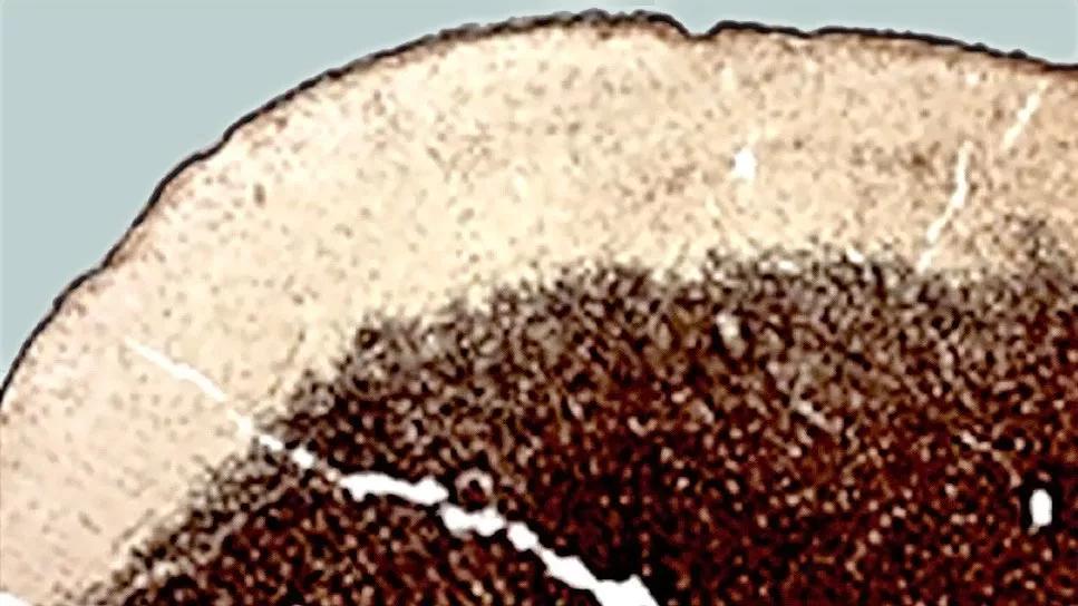
Image content: This image is available to view online.
View image online (https://assets.clevelandclinic.org/transform/208624b9-b167-4524-aa58-0ea5f3a410c4/gray-matter-lesions)
histology image of a gray matter lesion in a multiple sclerosis brain
In a first-of-kind analysis, Cleveland Clinic researchers have described how immune cells in the brain function differently at the edge of progressive multiple sclerosis (MS) lesions in white matter versus gray matter.
Advertisement
Cleveland Clinic is a non-profit academic medical center. Advertising on our site helps support our mission. We do not endorse non-Cleveland Clinic products or services. Policy
The Annals of Neurology publication (Epub 2024 Feb 12) reveals that microglia, immune cells that line the edge of demyelinated lesions in MS-affected brains, are proinflammatory/destructive in white matter but may be protective in gray matter. The findings, resulting from RNA sequencing of isolated brain tissue cells followed by immunocytochemistry, challenge the dogma that microglia are purely destructive in MS and only serve to fuel inflammation that expands the lesions in affected brains. Microglia may also play a role in slowing damage, depending on where they are located.
Understanding the different roles these cells play in progressive MS is essential for creating effective therapies that can reach the appropriate types of brain inflammation, says the study’s senior author, Bruce Trapp, PhD. Current treatments don’t directly address compartmentalized inflammation, or localized brain inflammation and damage behind the blood-brain barrier. The field is still learning about what causes this type of inflammation, a hallmark of progressive MS.
“The results indicate that a drug that suppresses microglial function is not a one-size-fits-all for progressive MS,” says Dr. Trapp, Chair of Cleveland Clinic’s Department of Neurosciences. “Some microglia may be destructive during MS while others may be protective. Loss of protective function may also play a role in disease progression. Reactivating certain functions of these cells with targeted therapies holds potential for treatment.”
Advertisement
Progressive MS lesions develop differently depending on their location in the brain. Lesions in white matter deeper in the brain are known to slowly expand, and microglia that surround these lesions are thought to play a role in this expansion. The role of microglia in the dynamics of gray matter lesion expansion are understudied because of limitations with imaging and preclinical models.
First author Anthony Chomyk developed a method to isolate microglia lining the lesions from white and gray matter and sequence their RNA to compare which genes were turned “on” or “off” between locations. Which genes are expressed can explain why different cells of the same type behave differently.
For this study, Chomyk analyzed donor brain samples from Cleveland Clinic’s MS rapid autopsy program. Dr. Trapp founded the program in 1994 to advance MS research imaging. It is now one of the largest rapid autopsy programs for MS in the world.
The study also provided insight that will help in detection of both types of lesions. Microglia on the edge of white matter lesions consume debris from demyelinated nerves, so the cells contain enough iron that it shows up in imaging.
Chomyk says that the study provides the bird’s-eye view needed for drug development. Investigators could explore the feasibility of targeting microglial function only on the edge of white matter lesions, for example.
“This study is another step forward in changing our field’s perception that all microglia have destructive roles in MS,” Dr. Trapp concludes. “To change the thought process in any field takes time, and additional data that support this change are needed. It’s difficult to change accepted perspectives, but the lack of effective therapies for progressive MS is telling us that we’ve missed something.”
Advertisement
Image at top shows a gray matter lesion with dense lines of microglia at the border. Courtesy of Chomyk et al., “Transcript profiles of microglia/macrophage cells at the borders of chronic active and subpial gray matter lesions in multiple sclerosis,” Annals of Neurology (Epub 2024 Feb 12), © The authors.
Advertisement
Advertisement

Two research projects aim to enable more personalized MS care in this population

Mixed results from phase 2 CALLIPER trial of novel dual-action compound

A co-author of the new recommendations shares the updates you need to know

Rebound risk is shaped by patient characteristics and mechanism of action of current DMT

First-of-kind prediction model demonstrates high consistency across internal and external validation

Real-world study also finds no significant rise in ocrelizumab-related risk with advanced age

Machine learning study associates discrete neuropsychological testing profiles with neurodegeneration

This MRI marker of inflammation can help differentiate MS from mimics early in the disease