Prevention of progression and complications is the primary goal

By Alexandra Villa-Forte, MD, MPH
Advertisement
Cleveland Clinic is a non-profit academic medical center. Advertising on our site helps support our mission. We do not endorse non-Cleveland Clinic products or services. Policy
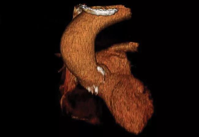
Takayasu’s arteritis (TAK) is an idiopathic granulomatous inflammatory disease that affects the aorta and its primary branches. The disorder is rare, mostly found in young women and is frequently chronic and relapsing. Physicians can encounter several diagnostic and therapeutic challenges when providing care for these patients.
One of the main challenges impacting patients with TAK is the difficulty in establishing early diagnosis. Diagnosis is often delayed for many years, and when patients are first identified, they often have irreversible arterial lesions. One reason for the delay in diagnosis is the absence of symptoms until vessel stenosis or occlusion are detected on physical exam or imaging studies. Another reason is the vague nature of symptoms that for years may be described by some patients as fatigue, arthralgias, myalgias, non-specific headaches or dizziness.
Later, symptoms suggestive of ischemia in a young patient, such as carotidynia, syncope or limb claudication lead to further evaluation with computed tomography angiography or magnetic resonance angiography, and arterial abnormalities are identified. These modalities are the primary diagnostic tools, along with positron emission tomography-computed tomography, though each modality has its strengths and shortcomings. Digital subtraction angiography is rarely needed for diagnosis.
Laboratory tests are not always helpful in establishing diagnosis or monitoring disease activity as the sedimentation rate (WSR) and C-reactive protein (CRP) may be normal at times of active disease, in up to 50% of patients.
Advertisement
Patients may also develop tachycardia with palpitation, chest pain, dyspnea, renovascular hypertension, mesenteric angina and less commonly, visual disturbances. On physical exam, absent or asymmetric arterial pulses and/or blood pressure, and vascular bruits may be noted.
While establishing early diagnosis is difficult, determining and monitoring disease activity in patients with TAK also proves challenging. Patients may be asymptomatic with normal WSR and/or CRP, and have evidence of a new arterial lesion, or they may have progression of a previous arterial lesion. Other patients may have elevated WSR and/or CRP without evidence of new or progressive arterial lesions.
Complications from arterial inflammation differ according to which vessels are affected. They include secondary hypertension, aortic regurgitation, congestive heart failure, aortic aneurysm, stroke and chronic limb claudication, among others. Vessel damage may occur not only from active inflammation but also from vessel fibrotic remodeling when the disease may be in remission. Patients with TAK are at risk of significant vascular morbidity over the years.
Risk assessment is very important in patients with TAK. The location and extent of arterial lesions should be carefully analyzed with an experienced radiologist to predict risks of major complications such as stroke, organ infarcts (e.g., kidney), progression of a thoracic aortic aneurysm, cardiac ischemia and severe limb functional disability. Risks from chronic immunosuppression should be incorporated in treatment decisions, especially in light of the young age of many patients with TAK, and all therapy-related risks should be clearly discussed with patients and families and closely monitored on a regular basis. Control of disease activity, halting vascular lesion progression and preventing complications are the primary goals of treatment.
Advertisement
TAK is a chronic or relapsing disease in the majority of patients and continuous or multiple courses of treatment are often needed. The care of these patients relies on close monitoring of disease over time by combining evaluation of symptoms, vascular physical exam, laboratory tests and sequential imaging tests. Imaging tests at regular intervals allow for evaluation of location and extent of arterial lesions and the identification of lesions in new vascular territories and progression of previous arterial lesions.
While TAK may be challenging to diagnose and treat, careful monitoring with the appropriate clinical expertise and tools and thoughtful, risk-sensitive treatment can improve outcomes for patients.
Dr. Villa-Forte is staff in the Center for Vasculitis Care and Research, Department of Rheumatologic and Immunologic Disease.
Advertisement
Advertisement

A conversation with Leonard Calabrese, DO

The case for continued vigilance, counseling and antivirals

High fevers, diffuse rashes pointed to an unexpected diagnosis

No-cost learning and CME credit are part of this webcast series

Summit broadens understanding of new therapies and disease management

Program empowers users with PsA to take charge of their mental well being

Nitric oxide plays a key role in vascular physiology

CAR T-cell therapy may offer reason for optimism that those with SLE can experience improvement in quality of life.