Locations:


Overlapping symptoms and complex treatment decisions make care coordination essential
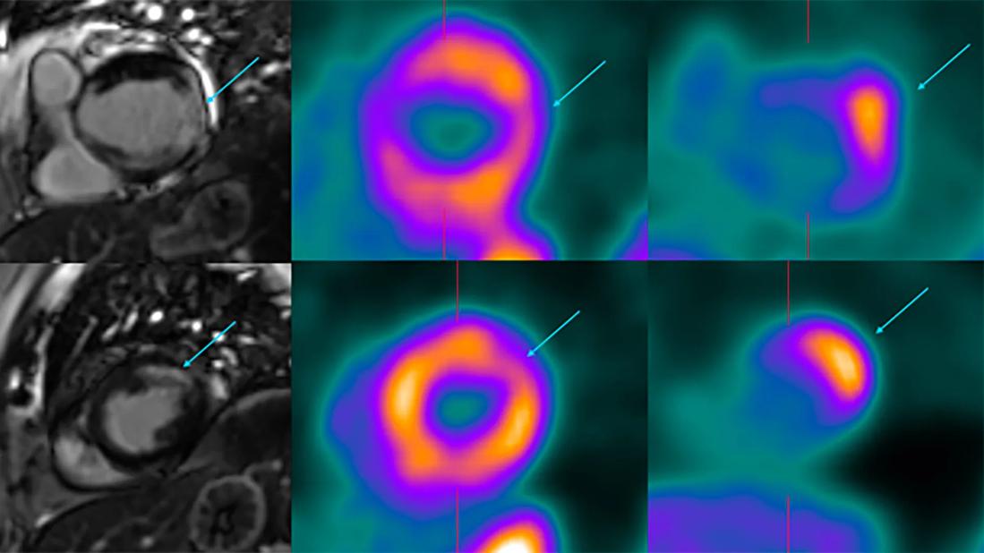
Image content: This image is available to view online.
View image online (https://assets.clevelandclinic.org/transform/b926689b-2281-418c-b5fd-45906068a3d9/isolated-cardiac-sarcoidosis)
six panels of MRI scans of the heart
Over the past decade, cardiac sarcoidosis (CS) diagnoses have risen dramatically. It remains unclear whether this increase is attributable to an increase in disease occurrence or to improved recognition by clinicians. Factors contributing to improved recognition include the development of advanced cardiac imaging techniques, a more standardized definition of the condition, and guidelines that help identify and treat patients with confirmed and suspected CS. However, CS is a complex and challenging condition that requires a multidisciplinary approach to treatment.
Advertisement
Cleveland Clinic is a non-profit academic medical center. Advertising on our site helps support our mission. We do not endorse non-Cleveland Clinic products or services. Policy
Sarcoidosis is a systemic disease, and its presenting signs and symptoms can be deceptively overlapping. Thus, multidisciplinary clinical assessment is critical to ensure optimal outcomes. Depending on presenting complaints, the patient’s journey at Cleveland Clinic can start with cardiac or pulmonary evaluation.
“At Cleveland Clinic, pulmonologists see almost every sarcoidosis patient, regardless of the combination of organ systems involved,” explains Brady Otten, MD, a pulmonologist at Cleveland Clinic. “Management decisions account for differing severity across organs, which requires coordination when different specialists may prioritize different treatment needs.”
Dr. Otten says he looks for a few things that suggest possible cardiac involvement in patients with sarcoidosis. Symptoms that can raise concern include palpitations, lightheadedness, dyspnea disproportionate to pulmonary findings and signs of heart failure.
“These symptoms are associated with several other cardiac-related conditions, which is what makes CS difficult to recognize,” says Dr. Otten. “Clinicians need to be thorough with their exclusion of other causes first.”
Particular attention should be paid to the occurrence of high-degree atrioventricular block in a young person or ventricular tachycardia of unclear etiology, explains Ziad Taimeh, MD, a cardiologist at Cleveland Clinic.
In patients with known sarcoidosis, Dr. Otten typically begins with symptom assessment and an electrocardiogram (ECG). Abnormal findings or concerning symptoms may prompt echocardiography, rhythm monitoring or referral to his colleagues in the Cardiac Sarcoidosis Center within Cleveland Clinic’s Heart, Vascular & Thoracic Institute, who specialize in diagnosing and managing CS. The most concerning clinical manifestations of CS include ventricular arrhythmias, heart block and heart failure.
Advertisement
A recent international Delphi study jointly led by Daniel Culver, DO, Chair of the Department of Pulmonary Medicine, and Christine Jellis, MD, PhD, MBA, Vice Chair of Operations for the Heart, Vascular & Thoracic Institute, provides a consensus from global experts on optimal diagnosis, monitoring and management of CS through the utilization of advanced cardiac imaging.
“This unique opportunity to bring together world experts from across multiple specialties and provide guidance on managing CS has helped set global expectations on how we should approach the management of these complex patients,” says Dr. Jellis.
Typically, most patients with pulmonary sarcoidosis or other forms of extracardiac sarcoidosis will have a screening ECG performed by their treating physicians, who are aware of the potential for cardiac involvement. If the ECG is abnormal or the patient has symptoms that cause concern for underlying CS, such as palpitations, dizziness or shortness of breath, then the next step may involve ordering an echocardiogram (echo) to assess cardiac structure and function. Concerning findings on echo include reduced cardiac systolic “pumping” function, thinning and aneurysmal deformation of the left ventricular wall, and other associated findings such as a pericardial effusion.
If the ECG, echo or symptoms are suspicious for CS, referral to a cardiologist with expertise in CS is recommended for additional evaluation. Imaging cardiologists, like Dr. Jellis and her colleague Bryan Abadie, MD, use advanced cardiac imaging techniques to help establish a probable diagnosis of CS.
Advertisement
Dr. Jellis describes imaging cardiologists as “similar to detectives who use advanced imaging tests like cardiac MRI and cardiac PET to uncover the true diagnosis.” Cardiac MRI allows not only assessment of cardiac function and structure but also detection of inflammation and scar via gadolinium contrast. A patchy pattern of contrast uptake, particularly in the left ventricular septum or the right ventricle, is highly suggestive of CS (Figure). Nevertheless, there is considerable overlap with other heart conditions, which can make the diagnosis challenging.
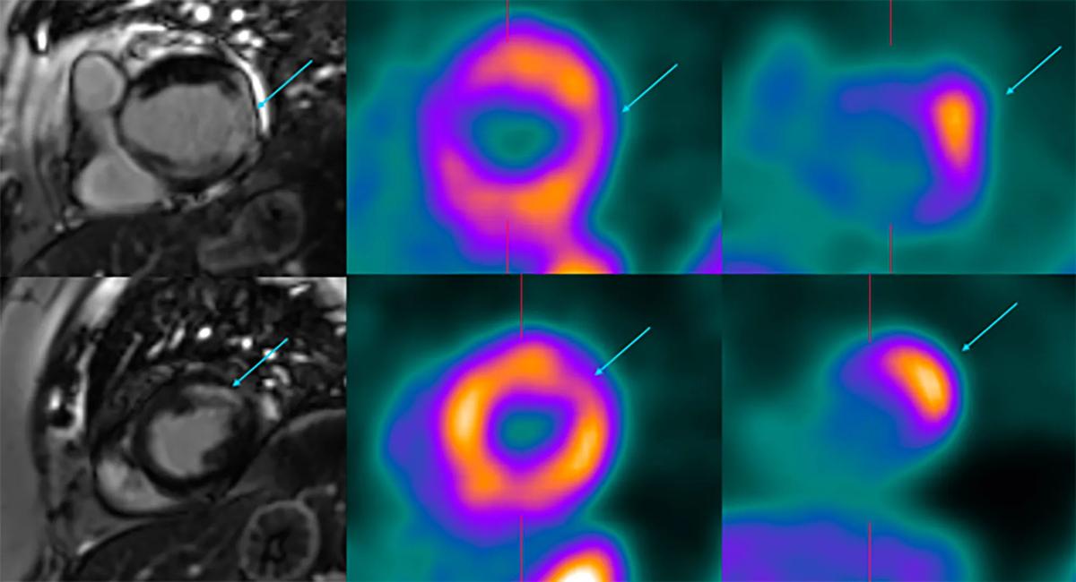
Image content: This image is available to view online.
View image online (https://assets.clevelandclinic.org/transform/b926689b-2281-418c-b5fd-45906068a3d9/isolated-cardiac-sarcoidosis)
Figure. Left: Cardiac MRI showing late gadolinium enhancement in a patchy distribution in the base (top) and mid (bottom) segments of the left ventricle. Middle: Corresponding areas of perfusion defect on cardiac PET. Right: Corresponding F18-fluorodeoxyglucose uptake. This “mismatch” pattern on cardiac PET is suggestive of active inflammation.
To help differentiate between active inflammation and scar tissue within the heart, cardiac PET imaging is often required. This unique imaging scan, performed by Dr. Abadie and his colleagues in nuclear cardiology, can help cement the diagnosis and monitor response to treatment. “Cardiac PET is a powerful test in our toolbox to diagnose and monitor sarcoidosis,” Dr. Abadie says. “It uses a nuclear tracer that resembles glucose to identify abnormal inflammation in the heart.”
While any tissue confirmation is highly recommended, it’s important to note that heart biopsy for CS may miss the diagnosis in about 75% of patients due to the patchy nature of the disease. “A negative heart biopsy result cannot exclude CS,” Dr. Jellis emphasizes. For this reason, if the patient has suspected sites of sarcoidosis outside the heart, such as enlarged lymph nodes in the chest, these are often better sites for biopsy. “When a patient undergoes PET imaging, our nuclear radiologists will image the entire body, not just the heart, to better identify safe places for biopsy,” she explains.
Advertisement
To complement the diagnosis, the cardiac team will arrange genetic testing to ensure the patient does not have a genetic inflammatory cardiomyopathy. Genetic inflammatory cardiomyopathies can have cardiac MRI and cardiac PET findings similar to those of CS and can commonly be mistaken for it.
Cleveland Clinic’s CS specialists also ensure there is adequate exclusion of coronary artery disease, typically through coronary angiography or coronary CT angiography. “An atypical presentation of a highly prevalent condition like coronary artery disease should not be missed in favor of a rare disease like CS,” Dr. Jellis notes.
Once CS is suspected or confirmed, management depends on how the disease is manifesting and whether there is evidence of active inflammation. Dr. Taimeh emphasizes that treatment should first focus on addressing the presenting cardiac issue, such as heart block, heart failure or arrhythmia.
Next, treatment should focus on suppressing any active myocardial inflammation present to improve heart function or prevent it from worsening. Patients presenting with ventricular arrhythmias or those with high-grade heart block will need a dual-chamber pacemaker/defibrillator, and those with heart failure due to reduced “pumping” function may require guideline-directed medical therapy for heart failure or even consideration for a left ventricular assist device (LVAD) or cardiac transplantation in severe cases.
“We have successfully transplanted patients with CS, with insignificant recurrence of cardiac inflammation,” explains Dr. Taimeh. “Although LVAD therapy remains a viable option, the risk of pump or driveline infection with immunosuppression remains high.”
Advertisement
Immunosuppressive therapy is typically considered when cardiac PET imaging shows active myocardial inflammation.
“When a patient has inflammation, the most universally effective medication is prednisone,” says Dr. Otten. “However, treating sarcoidosis with prednisone over an extended period can also be harmful to patients. It can cause decreased bone density, weight gain, increased blood pressure and blood sugar … the list goes on.”
He continues: “But if I’m seeing a patient who’s been diagnosed with CS and they have active inflammation, we’re going straight to prednisone along with a second immunosuppressive medication — typically, methotrexate, infliximab or other immunomodulators. The idea is that once that second medication sets in and is doing what it’s meant to do, we can slowly pull off the prednisone. Ideally, we want the patient off prednisone within several months so they’re just being treated with that immunosuppressive agent.”
He also notes that the decision to pursue steroid-sparing therapy must be based on a patient’s comorbidities, risk of side effects, route of administration and whether sarcoidosis is affecting other organs. In some cases, treatment decisions may be driven as much by disease in other organs as by cardiac findings. This is why a multidisciplinary approach to CS treatment can be so important.
In addition to coordinating care between pulmonary and cardiovascular medicine, specialists from ophthalmology, dermatology, neurology, hepatology and other areas are frequently involved in the care of patients with sarcoidosis, depending on whether a patient has manifestations of sarcoidosis outside the lungs and heart.
Cleveland Clinic’s multidisciplinary cardiac sarcoidosis team connects frequently and discusses their most complex cases at a twice-monthly cardiac sarcoidosis conference.
“Cardiac sarcoidosis is the greatest masquerader, and eyes with different lenses are often needed to capture the probability of occurrence,” says Dr. Taimeh.
“There’s a lot of collaboration and discussion around which diagnostic tests are needed,” Dr. Otten adds, “as well as whether the patient truly has CS, how the patient should be monitored and which treatments should be used.”
Dr. Abadie finds professional value in the meetings as well. “These multidisciplinary meetings also serve as a valuable way for our team to build professional relationships, develop research ideas and educate younger physicians about CS, all of which are important for improving care for our patients with CS.”
Because of its complexity, a multidisciplinary approach is central to managing CS effectively. CS is a difficult condition to diagnose since many clinicians may not frequently encounter it, and its symptoms can be seen in other, more-common conditions. However, when CS is missed, the consequences can be serious.
Abnormal findings on ECG or echo might suggest CS, but advanced cardiac imaging tests, such as cardiac MRI or PET, can strengthen the diagnosis. Management of these patients by cardiologists who are experienced in this area is key. They can advise on the need for defibrillators, in addition to managing heart failure in severe cases. If active CS is diagnosed, immunosuppression to suppress and resolve inflammation is important to prevent long-term damage to the heart.
“We’ve made headway in helping clinicians recognize that this is an impactful disease,” says Dr. Otten. “When it goes unrecognized, it can have severe effects on patients’ health, so awareness is important.”
Advertisement
How careful measurement, provocative testing and repeat assessment can maximize the diagnostic value of PAC

Cleveland Clinic experts discuss clinical clues, testing considerations and multidisciplinary care for patients with suspected short telomere syndromes
Cleveland Clinic’s new department chair discusses clinical priorities, prevention, stewardship and the evolving role of technology

A look at how physician work is valued and why survey participation matters

A look at the emerging link between intermittent hypoxia and broader health effects in COPD
Appropriate patient selection and clinician awareness remain key to broader use

Overexposure to the common alloy can lead to an autoimmune response called sensitization
An ICU pharmacist explains the impact of delirium on ICU patients and why there is a need for more research and improved screening tools
 Search