Offers recommendations on imaging and treatment across five clinical pericardial syndromes

A new international expert consensus document detailing the applications of multimodality cardiac imaging in the diagnosis, risk stratification, management and surveillance of pericardial diseases is filling a major education gap among physicians.
Advertisement
Cleveland Clinic is a non-profit academic medical center. Advertising on our site helps support our mission. We do not endorse non-Cleveland Clinic products or services. Policy
“There are no American clinical guidelines on this topic, and the latest European guidelines were published in 2015, before clinical trials of interleukin-1 blockers in recurrent pericarditis were conducted,” says Allan Klein, MD, Director of the Pericardial Diseases Center at Cleveland Clinic. “This document informs best practices with the goal of reducing variability in the evaluation and management of these patients.”
The document, published in JACC: Cardiovascular Imaging (2024;17[8]:937-988), was developed by an international committee chaired by Dr. Klein and comprising 22 leaders in the imaging and management of pericardial diseases. It provides disease-specific algorithms based on evidence supporting the use of echocardiography, cardiac computed tomography (CT) and cardiac magnetic resonance (CMR) to define disease severity and support earlier targeted treatment with interleukin-1 (IL-1) blockers.
Pericardial diseases have a wide variety of etiologies and are commonly seen in emergency departments, outpatient clinics, primary care offices and rheumatology practices. They are the second most common cardiac cause of chest pain after coronary artery disease, yet they are often misdiagnosed by clinicians.
“Viral/idiopathic etiology is most common,” says Dr. Klein. “However, post-cardiac injury — such as complications from cardiac surgery, arrhythmia ablation, device implantation, interventional cardiology procedures and the like — is increasing as a leading cause of acute and recurrent pericarditis. Other causes include autoimmune disease and malignancy.”
Advertisement
Echocardiography remains the first-line imaging modality for pericarditis due to its ability to assess pericardial effusion, heart chamber size and function, heart valves and hemodynamics in a portable, noninvasive manner at low cost. But the new document explains in exquisite detail when and how transthoracic echo should be employed, which conditions should prompt the use of transesophageal echo, and when CT or CMR should be used in place of, or as a complement to, echocardiography.
“Multimodality imaging, especially with CMR evaluation of pericardial inflammation and edema and echo evaluation of constrictive physiology, allows precise definition of disease presence and severity,” Dr. Klein says. “We can tell not only how bad the disease is but also predict approximately how many years it will take to achieve clinical remission and how many relapses the patient will have. We advocate for a new imaging-guided therapy approach for pericarditis.”
Because acute pericarditis is often undertreated, leading to recurrent disease, the ability to obtain information on disease severity in real time can promote earlier intervention and is expected to thereby improve patient outcomes. “With this approach, the timing of surgery — i.e., radical pericardiectomy — can be planned better,” notes Marijan Koprivanac, MD, MS, Surgical Director of the Pericardial Diseases Center. “Surgical success is greater when pericardial inflammation is the least, as there is less technical difficulty. MRI is especially helpful in determining the level of pericardial inflammation, along with other clinical tools.”
Advertisement

Image content: This image is available to view online.
View image online (https://assets.clevelandclinic.org/transform/da49e012-4b84-4cef-8e31-cadbf3e15110/MRI-showing-severe-pericaridal-inflammation)
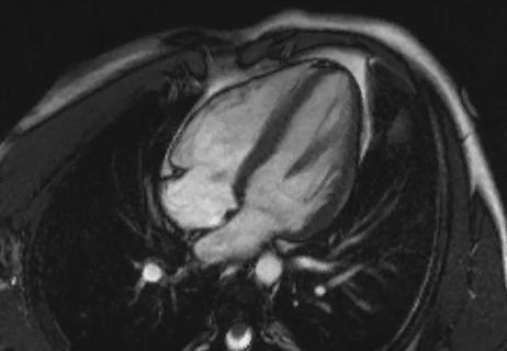
MRI showing severe pericardial inflammation on late gadolinium enhancement. The ability to see inflamed pericardium — "the halo effect" — on MRI is one of the most exciting examples of the value of CMR in staging pericarditis.
Complementary to the development of more precise imaging technologies is the advent of IL-1 blockers, including rilonacept, anakinra and the investigational therapy goflikicept. These biologic agents lessen the need for steroids and the accompanying risk of steroid dependence and its complications.
Dr. Klein led the pivotal RHAPSODY trial that first demonstrated the safety and efficacy of an IL-1 blocker in pericarditis in a randomized investigation (N Engl J Med. 2021;384:31-41). In this study of patients with pericarditis recurrence despite standard therapy, rilonacept was associated with rapid resolution of recurrent pericarditis episodes, allowing weaning from steroids and significantly reduced risk of subsequent recurrence compared with placebo. The results of RHAPSODY led to FDA approval of rilonacept.
In the new consensus document, the authors lay the groundwork for understanding pericardial diseases by discussing in detail the anatomy and pathophysiology of the pericardium and pericardial inflammation, while also explaining how biologic agents work.
The bulk of the document addresses five clinical syndromes: pericardial inflammation (acute/recurrent pericarditis), pericardial effusion, constrictive pericarditis, pericardial masses and congenital anomalies.
The role of multimodality imaging in each syndrome is discussed and outlined in one or several helpful tables. Recommendations are provided and key points highlighted. Also included are comprehensive algorithms for the diagnosis and management of acute and recurrent pericarditis, pericardial effusion and constrictive pericarditis (Figures 33-35, respectively).
Advertisement
In fact, Dr. Klein and his Cleveland Clinic cardiologist colleague Tom Kai Ming Wang, MBChB, MD, a vice chair of the writing committee, view the document’s abundance of quick-read summary tables, figures and algorithms as one of its key strengths. They point clinicians to the following specific examples for important takeaway guidance.
A table outlining the classification of pericardial diseases (Table 4), which includes two new subcategories of constrictive pericarditis, transient and effusive. “With transient constrictive pericarditis, constriction can come earlier in the disease course, but if the inflammation is properly treated with anti-inflammatories, the constriction can disappear,” Dr. Wang notes. “In effusive constrictive pericarditis, constriction is masked by effusion, but once the effusion is drained, then signs of constriction appear and should be treated aggressively to prevent progression to end-stage constrictive pericarditis.”
An imaging-guided approach to diagnosing and managing the spectrum of pericardial diseases. This includes inflammation and constriction as seen on CMR (Figure 8) and novel pericarditis severity grading criteria on CMR (Figure 9). “Constrictive physiology progresses along with inflammation,” Dr. Klein observes. “Treatment depends on where the patient is on the spectrum, and this figure allows quick visualization of the considerations.”
A table summarizing essentials of the therapeutic options for acute and recurrent pericarditis (Table 9). “This may be the most crucial piece of the document,” Dr. Klein says. “A patient who doesn’t respond to conventional therapy should go on IL-1 blockers before steroids or undergo pericardiectomy. Consider taking these steps early, because the conventional drugs have a high relapse rate.”
Advertisement
“Clinicians will find the suggested information on dosing, duration, tapering and monitoring of the various anti-inflammatory therapies for pericarditis very helpful, especially anti-IL-1 agents like rilonacept, which has only been FDA-approved for about three years,” Dr. Wang adds.
A simple algorithm for using imaging to diagnose constrictive pericarditis (Figure 23). “Constrictive pericarditis is often misdiagnosed because its symptoms mimic those of other diseases, such as heart failure,” Dr. Klein notes. “Although the usual treatment starts with diuretics and many patients ultimately need pericardiectomy surgery, medical therapy can be effective in transient constriction if it is caught early through optimal imaging.”
The consensus document notes that it has been endorsed by American College of Cardiology (ACC) Imaging Council and the Society of Cardiovascular Magnetic Resonance and should be used to inform the steps required for diagnosing and treating all forms of pericardial diseases.
Although a scarcity of clinical trial evidence prevented the document from being called a guideline, the growing number clinical trials being performed is expected to further refine clinical guidance. “We are now working with the American Heart Association on collaborative pathways and with the ACC on a concise clinical guideline,” Dr. Klein says.
This is good news for patients, who stand to benefit from increased knowledge among physicians. The writing committee hopes that early diagnosis and appropriate treatment will prevent many acute cases of pericarditis from becoming chronic or recurrent.
“Following the document’s imaging and treatment guidance should allow accurate early diagnosis and grading of pericardial diseases and eliminate the undertreatment of acute pericarditis, reducing the risk of debilitating chronic or recurrent disease,” Dr. Wang notes. “With colchicine (for 3-6 months) and NSAID (or high-dose aspirin) dual therapy being the preferred treatment for acute pericarditis and first recurrence, anti-IL-1 agents are preferred ahead of corticosteroids in subsequent recurrences or incessant pericarditis not responding to first-line therapy. The duration of these therapies is currently controversial but should continue at least 18 to 24 months. This should be followed by shared decision-making on whether to slowly wean or continue indefinitely, depending on clinical and imaging response and patient preference.”
Advertisement

A closer look at the impact on procedures and patient outcomes

Join us in Florida this winter for a long-standing CME favorite

Study finds important discrepancies among modalities and techniques

Study finds that in the absence of ACS, the yield is low and outcomes are unaffected

Study validates a noninvasive alternative to invasive coronary angiography

How Cleveland Clinic is using and testing TMVR systems and approaches

NIH-funded comparative trial will complete enrollment soon

How Cleveland Clinic is helping shape the evolution of M-TEER for secondary and primary MR