Locations:

Two case studies illustrate why it’s important to look at DXA data holistically when treating patients with fracture risk
Written by Susan Williams, MS, RD, MD, CCD, FACP, FACE; Leila Khan, MD; Angelo A. Licata, MD, PhD, FACP, FACE
Advertisement
Cleveland Clinic is a non-profit academic medical center. Advertising on our site helps support our mission. We do not endorse non-Cleveland Clinic products or services. Policy
Editor’s note: This is an abridged version of an article originally published in the Cleveland Clinic Journal of Medicine. The article in its entirety, including a complete list of references, can be found here.
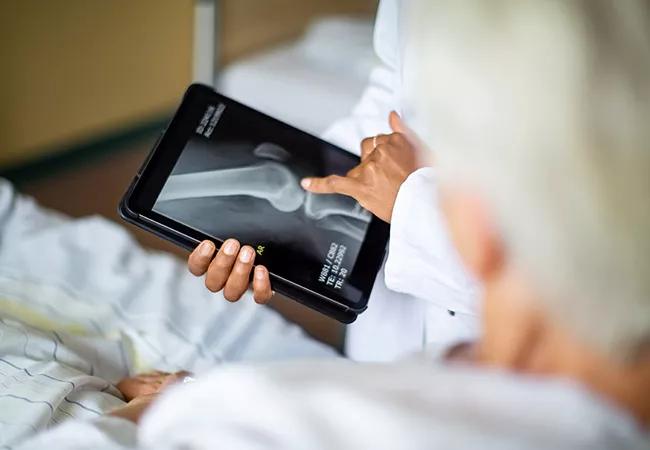
Our understanding of dual-energy x-ray absorptiometry (DXA) is evolving as new information emerges about skeletal qualities that contribute to bone strength apart from bone mineral density (BMD). Some of these characteristics are not detectable by DXA analysis. Hence, overdependence on DXA results, particularly for patient populations that the test was not designed for, may lead to poor clinical decisions.
DXA, originally developed for assessing fracture risk in postmenopausal women, is the gold standard test for diagnosing osteoporosis and monitoring its treatment. It can detect small but clinically relevant deficiencies in bone mass years before they are apparent on standard clinical radiographs, thereby allowing clinicians to intercede early to prevent fractures.
T-scores
DXA measures areal BMD (i.e., bone mineral content divided by the bone scanned area) in the spine, hip or forearm. Risk of fragility fracture is based on a calculated value called the T-score, which is the standard deviation of a patient’s measurement from the mean of a young, healthy reference population. Values and their significance are as follows:
Advertisement
As an assessment of fracture risk, T-scores are applicable only to untreated postmenopausal women and older men. Once drug therapy has started, T-scores do not accurately reflect risk.
Scientific advances have brought about a more complex understanding of the relationships between fracture risk, bone strength and bone density. T-scores do not always correlate with fracture risk or even with a patient’s history of fracture and hence can be misinterpreted, leading to inappropriate treatment recommendations. A T-score should not solely determine diagnosis and treatment and clinical data should appropriately modify the interpretation of results.
The following cases illustrate commonly encountered challenges if the DXA data and clinical presentation are incongruous.
A 35-year-old woman was referred for further guidance regarding a recent abnormal DXA test that had been ordered because of suspected “shin splints,” i.e., shin pain due to running. She was in good health, had been a recreational runner for decades with normal menstrual function and had a healthy diet with no tobacco or alcohol use. She had no history of fractures but reported that a maternal relative may have had osteoporosis. She said that all women in her family were petite, had small stature, and had low bone density. Her examination showed small skeletal structure but was otherwise normal. Blood tests were normal. Review of the recent DXA report revealed T-scores of-0.6 for spine and-2.5 for hip. The report also included “borderline osteoporosis” in the hip and recommended drug therapy. The patient was psychologically traumatized by this information and sought further guidance.
Advertisement
A 60-year-old woman underwent her first DXA test, resulting in a T-score of-1.5 for spine and a score of-2.0 for the femoral neck. Her physician was pleased that the scores were not in the osteoporotic range, since she had undergone surgical menopause 30 years earlier. The physician’s advice was to continue her healthy lifestyle, which included regular exercise, a vegan diet and daily calcium and vitamin D supplements. However, she had a radiologic and clinical diagnosis of spinal osteoarthritis, as well as bilateral wrist fractures from falls when she was in her 50s. Her family history included fractures in her mother and maternal grandmother. She used hormonal therapy after surgical menopause but stopped at age 45 due to concerns about risks and side effects. She has required no other prescription medication. Her blood test results were normal.
T-scores can be misinterpreted and lead to inappropriate treatment recommendations. Paradoxically, the patient in case 1 with a low hip T-score score may not be at immediate high risk of fracture, and a conservative approach may be reasonable if there are no significant risk factors, while the patient in case 2 with the osteopenic T-score is at very high fracture risk and requires aggressive therapy.
What are the reasons for this paradox? Advances in clinical science have revealed more complexities in the concepts of fracture risk, bone strength, and bone density. A T-score alone should not be the final arbiter of diagnosis and treatment. Clinical data can modify the interpretation.
Advertisement
The 35-year-old runner with a diagnosis of osteoporosis based on a low DXA hip T-score exemplifies a cascade of errors in fracture risk assessment. She may actually not be at immediate high risk of fracture, and a conservative approach may be reasonable.
The initial mistake was to order DXA for shin splints, which is not an indication for the test. This led to inappropriate use of T-scores, an incorrect diagnosis, prescription of a bisphosphonate in a healthy premenopausal woman with low fracture risk (and a chance of pregnancy) and unnecessary psychological turmoil for the patient.
Clinical factors are paramount in this case. The patient’s normal menstrual function implies sufficient estrogen production that should protect her skeleton without requiring additional medication. However, a low bone density may be suspicious for secondary problems and warrants a thorough family and clinical history to reveal possible causes. Laboratory testing would corroborate treatable options. A conundrum arises when no firm diagnosis can be found and the patient is healthy.
A diagnosis of borderline osteoporosis is contrary to guidelines of the International Society for Clinical Densitometry, and “low bone mass for age” is the preferred diagnostic term. Assessment with Z-scores rather than T-scores is appropriate for this healthy premenopausal woman. It is unclear whether her low bone density represents a loss of bone from a higher baseline, which may represent an underlying disease state, or whether she may have a genetic phenotype of low density despite having strong bone. Her family history and clinical examination are notable for small skeletal structure, suggestive of genetic inheritance of low bone mass. Studies of healthy premenopausal women with low bone mass show a spectrum of microarchitectural changes that mimic, to a minor degree, changes similar to osteoporosis. However, these women have a low risk of fracture. It is speculated that such architectural changes represent a pre-osteoporosis state.
Advertisement
A T-score indicating low bone density should not be ignored. Striving to maintain bone mass should be the guiding management principle, as she will enter menopause with a low bone mass and may experience fractures from estrogen deficiency earlier than expected for her age. A healthy lifestyle with adequate exercise and diet is the minimal therapeutic strategy. Attention should also be directed to any problems in menstrual function, eating, and hormonal disorders that may arise that would accelerate bone loss. However, skeletal pharmaceutical agents should not be considered unless evidence of bone fragility develops.
Surveillance of bone density with DXA is warranted, but a recommended interval has not been established in a premenopausal patient this young. In our practice, we consider every five to 10 years to be reasonable if no skeletal problems or illnesses arise.
In the case of the 60-year-old woman, the bone density report was misleading and discordant with the history of wrist fractures. Although vertebral and hip fractures attract the most attention from clinicians, wrist fractures also occur frequently and, unfortunately, are less likely to raise concern for osteoporosis evaluation and treatment. Data from a Medicare cohort showed only 7% of patients with wrist fractures have DXA testing within 6 months of such injuries, yet 20% later develop fractures of the hip or spine.
The American Association of Clinical Endocrinologists and the American College of Endocrinology point out that errors in DXA scan acquisition and analysis can affect interpretation, and they encourage clinicians to review actual scan images and data rather than rely solely on a report, especially when it is discordant with the clinical picture. Assuming this patient’s DXA report was of good quality, there are other reasons to question whether it reflects actual bone risk. Spinal arthritis can cause spinal DXA to be falsely normal and obscure bone deficiency. In women with a history of fracture, trabecular bone score technology usually reveals abnormal bone even when T scores are normal.
This patient’s multiple risk factors (i.e., age, history of fractures and estrogen deficiency since stopping hormone replacement after hysterectomy) attest to a weakened skeleton from osteoporosis, and a T-score that meets the osteoporosis threshold is not required to begin pharmaceutical treatment.
Further, advising only the use of calcium and vitamin D is inadequate management. Her provider should recommend that she use an antiresorption agent as first-line therapy and consider anabolic drugs if there are problems with the initial drug choice. She should not reinstate hormone therapy at her age for bone health alone as there may be increased risk for cardiovascular disease. However, this caveat is not absolute and requires a balance of risk and reward if hormone therapy is also needed for vasomotor, genitourinary or other problems.
Clinicians can expect to see patients similar to those in these two cases. Population data indicate that 2.5% of premenopausal women have T-scores below the reference range, and 6% of patients with any fracture have normal BMD tests. We recommend the following approach:
Assess patients using DXA and FRAX
These are still the major tools for assessing fracture risk. However, their results should not be regarded as absolute. The practitioner, not the technology, is the final arbiter for diagnosing disease.
Use T-scores as a guide for postmenopausal women and older men
A T-score of less than -2.5 is the intervention threshold for diagnosing and treating osteoporosis. However, keep in mind that patients with spine or hip T-scores in the normal or osteopenic range may require treatment for osteoporosis if the clinical history shows fractures. In such cases, one should not wait for T-scores to reach the critical -2.5 before intervening.
Use Z-scores for premenopausal women and young patients of both sexes. A low bone Z-score in healthy men or women indicates a generally low risk for fracture and is adequately treated with good nutrition, exercise, healthy lifestyle and skeletal surveillance. In the presence of fractures, illness that might affect the skeleton, or other risk factors, a more aggressive therapeutic approach may be indicated.
Advertisement

Pheochromocytoma case underscores the value in considering atypical presentations

Advocacy group underscores need for multidisciplinary expertise

A reconcilable divorce

A review of the latest evidence about purported side effects

High-volume surgery center can make a difference

Advancements in equipment and technology drive the use of HCL therapy for pregnant women with T1D

Patients spent less time in the hospital and no tumors were missed

A new study shows that an AI-enabled bundled system of sensors and coaching reduced A1C with fewer medications