Locations:

Motion-tracking Brillouin microscopy pinpoints corneal weakness in the anterior stroma

Image content: This image is available to view online.
View image online (https://assets.clevelandclinic.org/transform/65eab779-9f0e-4e4d-958f-8bb98ec261ea/keratoconus-eye)
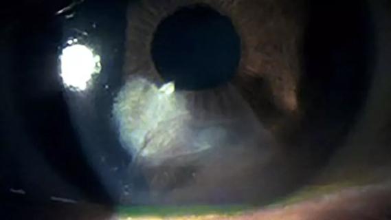
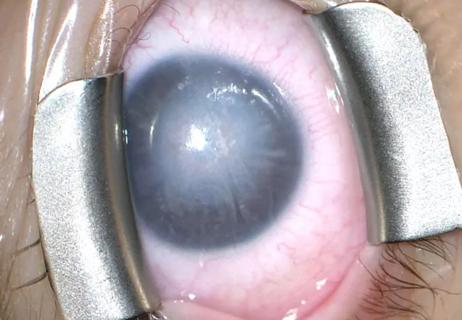
An eye with keratoconus
Advertisement
Cleveland Clinic is a non-profit academic medical center. Advertising on our site helps support our mission. We do not endorse non-Cleveland Clinic products or services. Policy
Up to 5% of people worldwide have keratoconus. Treating them with corneal cross-linking as early as possible helps stop the progression of permanent vision loss. Unfortunately, there’s no clear protocol for early detection of the disease.
We can perform various assessments and gather imaging data — with a slit lamp, corneal topography and multimodal imaging — but sometimes all that information is more confusing than clarifying, especially in subclinical keratoconus.
It’s thought that an underlying mechanical defect precedes keratoconus development. Epithelial remodeling and anterior and posterior surface elevation have been proposed as early indicators. Corneal curvature and thickness also could be indicative.
So, even with all of those metrics in the age of artificial intelligence, why are we still having difficulty identifying patients with subclinical keratoconus? It’s because none of those metrics are effective for detecting the earliest stages of the disease.
In a systematic review of subclinical and forme fruste keratoconus, most of the corneas appeared normal on slit lamp and had normal topography.
The KISA% index, an algorithm based on topography where 0-60 is considered normal and 100 or higher is considered keratoconus, also isn’t foolproof. In a study of Cleveland Clinic Cole Eye Institute patients with progressive keratoconus, we found that 20% of the diseased eyes were misclassified by KISA%, scoring in the normal range. Other research also has shown that KISA% lacks sensitivity.
Advertisement
Corneal epithelial thickness has been thought to partially or totally mask irregular corneal curvature. If that’s the case, we should be able to see changes in early stages of keratoconus on epithelial maps. However, a 2025 Cole Eye Institute study found no demonstrable difference between the epithelial maps of normal eyes and those with subclinical or manifest keratoconus. There was no difference in any parameter, location or metric. What’s more, epithelial mapping has never once performed well in studies of eyes with early keratoconus that still have normal corneal topography.
A global consensus on keratoconus was published in 2015. It included a very straightforward but controversial statement that posterior elevation abnormalities must be present to diagnose subclinical keratoconus. So, in another 2025 Cole Eye Institute study, we evaluated that statement. We screened over 1,200 papers and found 29 that compared the ability of posterior corneal surface, anterior corneal surface or corneal thickness to identify subclinical keratoconus. We found that less than 14% of that published literature reported posterior elevation or posterior surface changes as the best indicator of the disease. Even when assessed by time period, there was never a range of years in which studies reported posterior surface as better than anterior surface or corneal thickness at differentiating keratoconic eyes.
Theoretically, we should first see evidence of keratoconus in corneal biomechanics.
Brillouin microscopy imaging is a newer technique to assess corneal biomechanics. It involves shooting beams of light into specific points of the cornea. Shifts in light frequency, measured by spectrometer, indicate the stiffness and strength of corneal tissue at those points.
Advertisement
In an early ex vivo study using Brillouin imaging, researchers learned that keratoconic tissue was significantly softer and weaker in the cone, although tissue outside the cone was the same as in healthy corneas. It was the first proof of focal weakening in keratoconus.
Now at the Cole Eye Institute, we have developed motion-tracking Brillouin microscopy. It’s different from commercially available Brillouin imaging in that it incorporates OCT and ocular tracking. A 2020 in vivo study found that corneas with keratoconus were weaker in the anterior stroma compared with healthy corneas. So, using that information, we now are using motion-tracking Brillouin microscopy to gather data from the anterior cornea and convert it into two-dimensional, color-coded maps.
Our first study of motion-tracking Brillouin microscopy, in 2023, found distinct differences among normal, keratoconic and laser-corrected corneas. While normal anterior corneas were uniformly stiff, there was some loss of stiffness in the laser-corrected corneas and a real focal weakening in the cones. There were no differences in the periphery, however.
Our 2024 study found that motion-tracking Brillouin microscopy could completely differentiate corneas with subclinical keratoconus from healthy corneas. Brillouin imaging outperformed all Scheimpflug tomography metrics. The differences in the focal profiles of these eyes were immediately evident in the Brillouin shift maps.

Image content: This image is available to view online.
View image online (https://assets.clevelandclinic.org/transform/445296bd-b835-438e-bc7a-1b6b99426061/brillouin-mapping-subclinical-keratoconus)
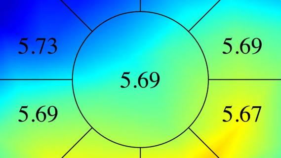
Motion-tracking Brillouin maps.
In 2025, we used motion-tracking Brillouin microscopy to study anterior, middle and posterior depths of corneas with early, subclinical or no keratoconus. Differences in corneal strength were most pronounced in the anterior stroma in eyes with keratoconus and least pronounced in the posterior stroma.
Advertisement
As we’ve studied this data, we’ve noted that the weakest point of the cornea seems to closely correspond with the thinnest point. Even in a normal cornea, that’s where the softest point is. In another 2025 study, when we plotted softest and thinnest points of corneas, we could clearly identify eyes with subclinical and early keratoconus by looking at their mechanical profile within this narrow region of the cornea.
All of this tells us where to look for keratoconus development. We now know where those earliest mechanical changes will be evident. They will be localized to the anterior third of the anterior stroma, specifically at the thinnest corneal point.
We will become even more precise over time, but already we can significantly reduce the area that we need to scan to detect subclinical keratoconus. The more we learn about the underlying mechanics of the disease, the earlier we will be able to treat patients.
Dr. Randleman is Co-Director of Refractive Surgery at Cleveland Clinic Cole Eye Institute. This article was based on his booth presentation at the 2025 American Academy of Ophthalmology meeting.
Advertisement
Advertisement

Study reveals more about the pathophysiology of Salzmann’s nodular degeneration
Studies continue to indicate effectiveness and safety
Identifies weak spots in the cornea before shape change occurs
Novel use of tPA reduces fibrin response in the eye

Researcher gives guidance for clinical trials

How computational tools and personalized biomechanics can improve keratoconus detection, ectasia risk assessment and surgical outcomes

Untreated seropositive erosive RA led to peripheral ulcerative keratitis

The drug does not provide lasting disease control for most patients