Locations:

How to screen for and manage treatment-triggered uveitis
Image content: This image is available to view online.
View image online (https://assets.clevelandclinic.org/transform/637e26df-5a56-4f70-bbb6-d2f2e7d208ff/23-EYE-3929962-CQD-Inflammatory-complications-clinical-trials_jpg)
23-EYE-3929962-CQD-Inflammatory-complications-clinical-trials
Inflammation in the eye (uveitis) is somewhat expected due to ocular surgery or infection. But when it’s unexpected — such as following an anti-VEGF injection in a patient with age-related macular degeneration (AMD) — it can be troubling, says Sunil Srivastava, MD, a vitreoretinal disease and uveitis specialist at Cleveland Clinic Cole Eye Institute.
Advertisement
Cleveland Clinic is a non-profit academic medical center. Advertising on our site helps support our mission. We do not endorse non-Cleveland Clinic products or services. Policy
“I think it strikes fear in most ophthalmologists,” he says. “It causes us to wonder what we’re missing and if the patient is at risk of permanent vision loss.”
Dr. Srivastava presented on inflammatory complications in clinical trials at the 2023 Cole Eye Institute Retina Summit in New Orleans, a prelude to the 2023 Association for Research in Vision and Ophthalmology (ARVO) annual meeting.
In the last few years, inflammatory complications have been seen in several products: brolucizumab, abicipar, pegcetacoplan and faricimab, to name a few.
“We all heard rumors of inflammation in late 2019. There was even a hint of it in clinical trials,” says Dr. Srivastava. “By early 2020, there were social media posts about a concerning case involving brolucizumab. That one case turned into several cases. Then ASRS [American Society of Retina Specialists] sent out a safety update, stating that they had received reports of inflammation following brolucizumab injection but that the etiology was unclear, and long-term outcomes and optimal treatment strategies were not yet defined.”
At that point, there had been 14 cases of retinal vasculitis, 11 of which were occlusive. In an August 2020 press release, the drug manufacturer stated the rate of retinal vasculitis or vascular occlusion was 4.71 per 10,000 injections. The manufacturer then convened a safety review committee.
“As we reviewed the cases, looking carefully at imaging, we realized that the rate of inflammation likely was higher than initially described,” says Dr. Srivastava, a member of the safety review committee.
Advertisement
Additionally, the committee found that patients developing inflammation had a 16% risk of mild vision loss (losing more than 15 letters) and a 10% risk of severe vision loss (losing more than 30 letters). Inflammation could happen at any time — not just in the first few months after treatment, but even 12, 14 or 18 months later.
“My take on this is that inflammation likely was present on imaging but not recognized, and retreatment occurred,” says Dr. Srivastava. “Inflammation or retinal occlusive disease could erupt after multiple treatments, months later, indicating that it was a cumulative issue in some eyes.”
What does this mean to ophthalmologists? Dr. Srivastava offers three key insights:
1. Inflammation is difficult to assess. “A slit lamp in a retina office is sometimes no better than a pen light,” he says. “Most of the time, it’s not as bright as we need. Additionally, office exams are quick. It’s easy to miss keratic precipitates and inflammatory cells.”
Imaging may not be reviewed closely, he adds. Even in a reading center, a patient’s OCT, fundus photo and fluorescein angiogram won’t be reviewed at the same time.
“In a uveitis clinic, we study all those images together, looking for subtle signs of inflammation and changes over time,” says Dr. Srivastava. “We also use clinical data to help us determine what’s happening with the patient. The reading center doesn’t have this data because the patients are masked.”
2. Inflammation may occur more in the real world than in controlled clinical trials. “Clinical trial patients are typically healthy ‘ideal’ patients,” says Dr. Srivastava. “More patients in the real world will be primed for inflammation. They may be sicker. They may have a history of systemic inflammatory disease or surgery, or have had previous treatments. They may be on medications that promote inflammatory disease.”
Advertisement
3. Inflammatory complications can present months after beginning therapy. Because inflammation is threatening to a patient’s vision, ophthalmologists have cause to hesitate before administering additional or new medication.
These points also apply to gene therapy trials, where inflammation is prevalent regardless of injection location or vector type. (Patients with retinal dystrophy and diabetic retinopathy likely already have inflammatory cells visible on clinical exam, even before receiving gene therapy.)
Ocular inflammation is seen in almost every gene therapy animal study, according to Dr. Srivastava. The degree of inflammation is related to dose. Histologic changes and cellular infiltration occur even in the absence of clinically apparent inflammation. And the effect of immunosuppressive pretreatment varies widely.
Considering the link between injectable therapies and inflammation, ophthalmologists can take steps to protect their patients. Dr. Srivastava offers these tips:
Advertisement
“Inflammation is a known complication of all drug therapies, although it is not completely understood,” concludes Dr. Srivastava. “It’s easy to miss subtle inflammation. We need to carefully examine eyes and image them longitudinally. Severe vision loss is possible once inflammation starts, so we must address it aggressively.”
Advertisement
Advertisement

The advanced stage of diabetic retinopathy is among the most challenging for retinal surgeons
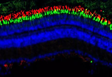
Researchers to study retinal regeneration in zebrafish with new grant from National Eye Institute
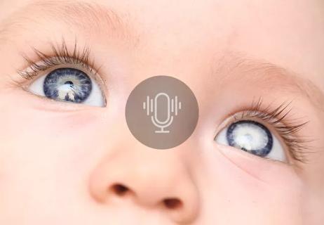
Leukocoria is the most common symptom of this rare childhood eye cancer

The Center for Environment, Place and Health Research connects clinicians with mapping tools and other real-world data resources to help guide medical insights

A scannable recap of recent volumes and clinical metrics from Cleveland Clinic

The drug does not provide lasting disease control for most patients

Nurse manager turns a patient safety concern into a data-driven effort to improve protocols and inspire innovation

Nurse draws on frontline experience and an engineering background to develop a new approach to incontinence management