Locations:


From Pavlik harness to periacetabular osteotomy, treatment approach varies by age and stage
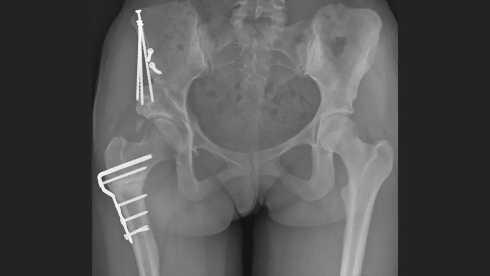
Image content: This image is available to view online.
View image online (https://assets.clevelandclinic.org/transform/12c03ef5-e27a-4af3-a1b7-c1a5ffe37a13/9189459-periacetabular-osteotomy-feature)
An X-ray of a patient's hips, showing opaque bones with bright white pins in them
Hip dysplasia can look very different in an infant than it does in a teenager or young adult, and the timing of the diagnosis can shape the treatment approach.
Advertisement
Cleveland Clinic is a non-profit academic medical center. Advertising on our site helps support our mission. We do not endorse non-Cleveland Clinic products or services. Policy
For clinicians, understanding how diagnosis and management evolve with age can help ensure timely referral and appropriate imaging.
In this article, David Gurd, MD, a Cleveland Clinic pediatric orthopaedic surgeon, explains how hip dysplasia is identified and treated across different stages of development.
“Hip dysplasia is not always indicated by hip pain,” Dr. Gurd says. “Infants and even young children who are walking typically do not have pain. Pain doesn’t become a primary complaint until adolescence, particularly when kids start playing school sports.”
Regardless of symptoms, the earlier the diagnosis, the better, he notes. Undiagnosed hip dysplasia may lead to pain later in life, as well as activity limitations, arthritis and hip replacement.
Diagnosing hip dysplasia in infancy starts with knowing which babies warrant special evaluation. Risk factors for hip dysplasia include:
Physical examination may include Barlow, Ortolani and Galeazzi testing, depending on the patient’s age.
When the examination or risk factors raise concern, ultrasound is the preferred imaging screening during the first several months of life.
X-rays become increasingly important for diagnosing hip dysplasia as children get older.
“As children age, we’re more likely to order X-rays when they develop soreness with walking or sports,” Dr. Gurd says. “That’s often how previously undiagnosed hip dysplasia is discovered.”
In older patients, hip dysplasia can be uncovered during the evaluation of labral tears, sometimes following a failed or recurrent repair. In these patients, the labral tear may be only part of the problem. Underlying dysplasia can continue to overload the labrum, so identifying the structural cause is essential before deciding on treatment.
Advertisement
“From infancy through young adulthood, hip dysplasia is really a different condition at each stage,” Dr. Gurd says. “As patients grow, both the way we detect it and the options we have to treat it change dramatically.”
When hip dysplasia is identified early, treatment is often relatively straightforward. Infants with a dislocated hip can frequently be treated with a Pavlik harness, which helps keep the hips in an abducted position and prevents adduction, encouraging the femoral head to remain seated within the socket as the joint develops.
Once the femoral head is back in the socket, the harness or a hip abduction brace helps keep it there while the socket continues to mature.
“The earlier we find a dislocated hip, the more likely we are to correct it with a brace rather than an operation,” Dr. Gurd says. “Early treatment can make a tremendous difference in how much intervention a child needs.”
When diagnosis occurs later in infancy, treatment often becomes more involved. By ages 6 to 14 months, a dislocated hip is less likely to respond to bracing alone, and many children require surgical reduction. Following hip reduction, a spica cast is typically used to maintain stability while the joint remodels.
Follow-up imaging helps the care team confirm that the hip is staying in place and the socket is developing as expected. Additional casting and subsequent bracing may be required.
“The longer we can keep the femoral head centered in the socket, the more opportunity the socket has to develop normally,” Dr. Gurd says.
Advertisement
When hip dysplasia is not diagnosed until a child has been walking for some time, treatment becomes considerably more complex. Closed reduction may no longer be feasible, and attempting to force the femoral head into the socket can increase the risk of avascular necrosis.
These patients frequently require open reduction surgery. Surgeons must first address the structures preventing the femoral head from returning to the socket, including tight soft tissues, an elongated ligament and tissue within the acetabulum itself.
To avoid excessive pressure on the hip and reduce the risk of avascular necrosis, a femoral shortening osteotomy is often performed, allowing the hip to be reduced more gently.
Depending on the child’s anatomy and the severity of the dysplasia, surgeons may also reshape the pelvis with procedures such as a Salter or Dega osteotomy to improve coverage and help keep the hip stable.
“By this age, we’re no longer talking about a brace,” Dr. Gurd says. “We’re often talking about a much bigger reconstructive operation.”
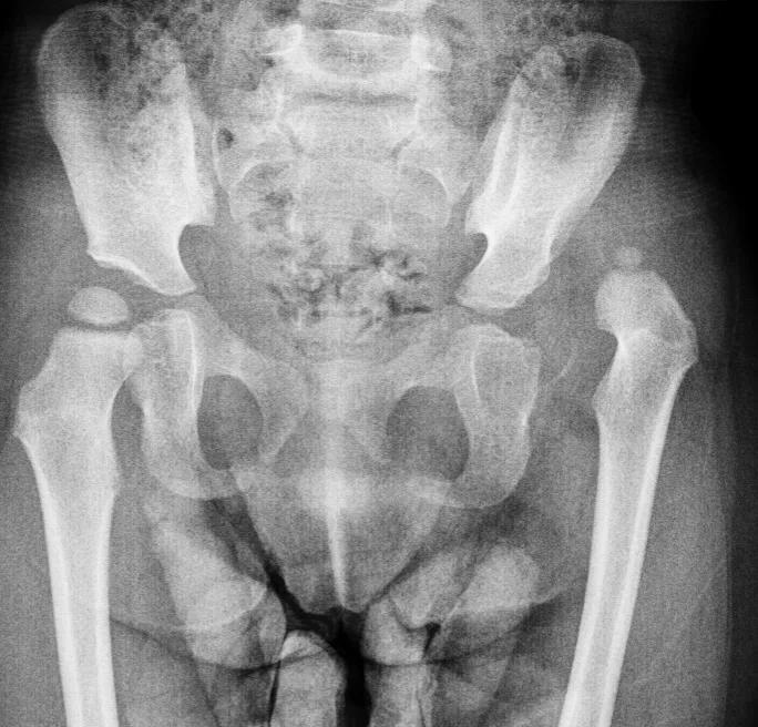
Image content: This image is available to view online.
View image online (https://assets.clevelandclinic.org/transform/822faa35-05a0-4841-a353-e8324bf593c5/9189459-left-hip-dislocation)
Left hip dislocation in a 2-year-old patient.
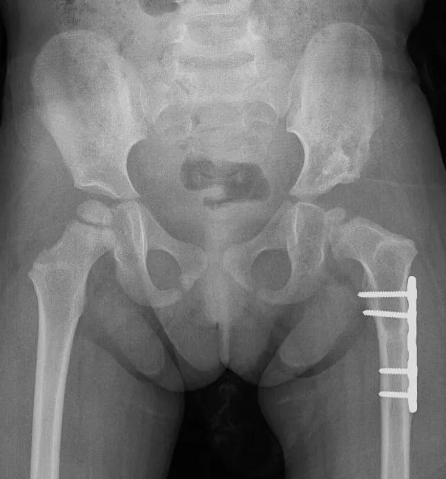
Image content: This image is available to view online.
View image online (https://assets.clevelandclinic.org/transform/2ea3dd7c-693d-4ffa-8256-5f6fd9b95317/9189459-open-reduction-femoral-shortening)
After open reduction and femoral shortening osteotomy to relocate the hip to the correct position.
In adolescents and young adults, the hip may stay in the socket but still lack adequate coverage. That changes how forces move through the joint and can lead to labral tears, pain and early cartilage damage.
By this stage, patients are no longer candidates for the pelvic osteotomies used in younger children because skeletal growth is complete. Instead, treatment may require a periacetabular osteotomy, a specialized hip preservation procedure designed to improve coverage of the femoral head.
Advertisement
During the procedure, surgeons make precise cuts through portions of the ischium, pubis and ilium to free the socket. They can then rotate it into a better position over the femoral head and secure it with screws.
“Periacetabular osteotomy is a very specialized operation that requires a careful balance,” Dr. Gurd says. “Too little correction leaves the hip undercovered, while too much can create femoroacetabular impingement. The goal is to achieve more normal joint mechanics, reduce stress on the cartilage and labrum, and preserve the native hip for as long as possible.”
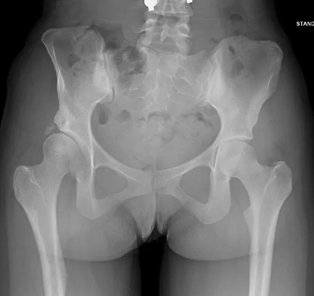
Image content: This image is available to view online.
View image online (https://assets.clevelandclinic.org/transform/b2155088-e8b9-4892-adb8-a11bfe8f4bec/9189459-acetabular-dysplasia)
Acetabular dysplasia with a shallow acetabulum on the right, leading to instability of the right hip in a 21-year-old patient.
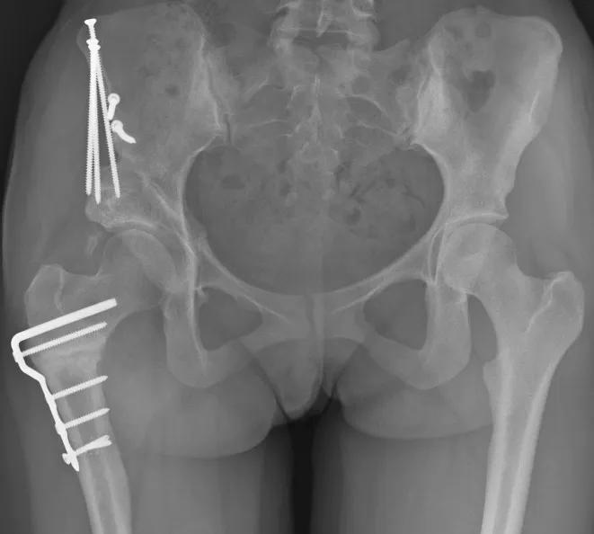
Image content: This image is available to view online.
View image online (https://assets.clevelandclinic.org/transform/5a34b800-2674-4620-ad74-39d9f2f765e4/9189459-periacetabular-osteotomy)
After periacetabular osteotomy and derotational osteotomy of the right femur to provide better coverage of the hip joint.
Treatment may change with age, but the goal remains the same: to create a stable, functional hip and prevent or delay arthritis. According to Dr. Gurd, outcomes are generally excellent when a reduced hip can be restored and maintained, whether through bracing in infancy or reconstructive procedures later.
Outcomes following periacetabular osteotomy can be highly favorable, particularly when patients are treated before substantial cartilage damage develops. While long-term data are still emerging, studies have shown that many patients continue to do well 10 to 20 years after this surgery. For many patients, success is measured not by eliminating hip replacement, but by delaying it for decades.
“Hip replacement is an excellent operation, but it doesn’t last forever,” Dr. Gurd says. “If we can preserve a patient’s native hip and delay a replacement from their 30s or 40s until their 60s or 70s, that’s a huge win.”
Because hip dysplasia presents differently at every stage of development, maintaining a high index of suspicion is important. Whether evaluating an infant with risk factors or an adolescent with persistent hip pain, timely imaging and referral can substantially influence treatment options and long-term outcomes.
Advertisement
“The quicker we identify and treat hip dysplasia, the less surgery a patient is likely to need and the better they’re likely to do,” Dr. Gurd says. “No matter the patient’s age, it’s important to keep dysplasia on your radar.”
Advertisement
Percutaneous stabilization can increase mobility without disrupting cancer treatment

Study shows that postop function is closer to normal than with total hip arthroplasty

Study challenges assumptions about risk evaluation in total hip revision

Study highlights the need for objective functional measures as value-based care expands

Reduces likelihood of achieving CMS-defined substantial clinical benefit at one year

Recognition could help guide health policy, workforce development and resource allocation worldwide
How it’s similar but different from the direct anterior approach
Should surgeons forgo posterior and lateral approaches?
 Search