Locations:

Case study of radial-to-axillary nerve transfer for tumor-related deltoid nerve injury
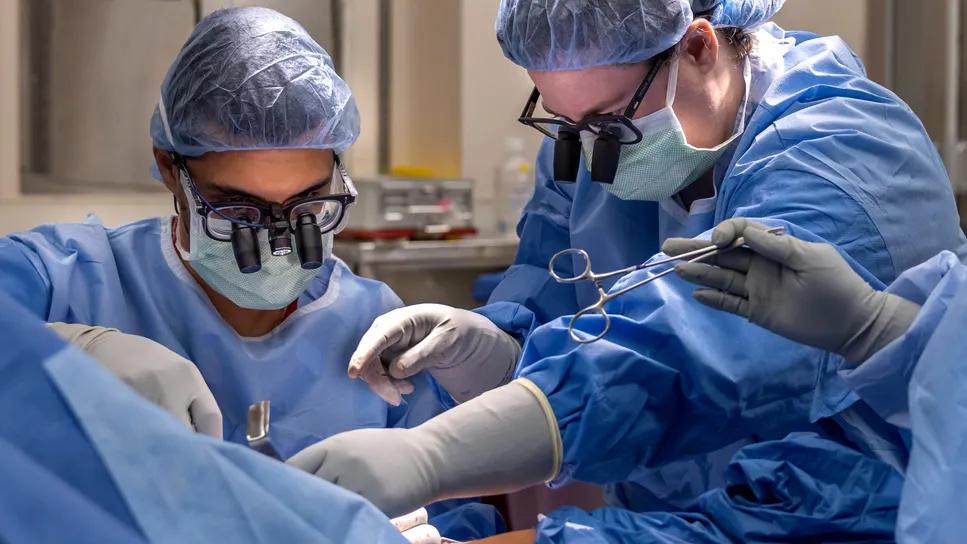
Image content: This image is available to view online.
View image online (https://assets.clevelandclinic.org/transform/29ea56e4-97fd-4f8d-b125-c2e5bbedf13d/nerve-transfer-after-ewing-sarcoma-therapy)
two surgeons performing an operation
In April 2025, a formerly athletic 14-year-old male with loss of function in his right deltoid secondary to Ewing sarcoma presented to Cleveland Clinic peripheral nerve neurosurgeon Megan Jack, MD, PhD, for evaluation.
Advertisement
Cleveland Clinic is a non-profit academic medical center. Advertising on our site helps support our mission. We do not endorse non-Cleveland Clinic products or services. Policy
Five months earlier, Dr. Jack had performed an open biopsy of a mass on the patient’s neck, which led to his cancer diagnosis. The tumor had been discovered on MRI, which was performed after the boy’s persistent shoulder pain failed to respond to treatment for presumed tendinitis.
The patient underwent 14 cycles of chemotherapy with vincristine/doxorubicin/cyclophosphamide plus ifosfamide/etoposide for the Ewing sarcoma, supplemented by radiation therapy initiated during the latter half of the chemotherapy course.
Throughout his chemotherapy, the patient’s nerve function was monitored closely by Dr. Jack with electromyography (EMG). “The tumor was wrapped around two exiting nerve roots, which is why his condition presented as pain in the shoulder,” Dr. Jack explains. “We were watching to see if tumor shrinkage from the cancer treatment would take enough pressure off the affected nerves to allow them to heal on their own.”
The tumor responded well to the treatment, and repeat EMG confirmed that the patient’s biceps function was gradually returning to normal. At five to six months into his treatment, however, his deltoid function lagged, prompting his latest presentation to Dr. Jack for evaluation for nerve transfer surgery.
Physical examination by Dr. Jack confirmed the near-complete return of function in the patient’s right biceps, but not in his right deltoid. EMG was once again performed and confirmed the lack of response to electrical stimuli in the muscle.
“We had a short window before the end of the patient’s radiation therapy, during which time it would be optimal to perform a nerve transfer,” Dr. Jack says. “It was our hope that we could restore the deltoid function and he could get back to playing the sports he enjoyed — basketball, football and lacrosse.”
Advertisement
In May 2025, Dr. Jack performed a radial-to-axillary nerve transfer. An incision was made via a longitudinal, posterior approach to the proximal humerus. Dissection was performed to separate the brachial fascia from the triceps muscle, and the radial nerve and profunda were visualized. “Our initial goal with this surgery was to find the axillary nerve, go down within the quadrilateral space, and decompress the nerve,” says Dr. Jack. “We also did intraoperative testing to make sure that when we stimulated the nerve, we didn’t see any motor recovery.”
Next, the branch of the radial nerve that supplies the triceps extension but does not contribute to wrist or digital extension was identified and dissected proximally to the inferior border of the teres major, and the fascia overlying the inferior third of the teres major was released. Then the isolated branch of the nerve was coapted to the axillary nerve in end-to-end fashion, using 8-0 nylon sutures. The fascia and subcutaneous and skin layers were closed to complete the procedure.
The patient remained in the hospital overnight for observation and pain control. Before discharge the next day, he was counseled about the need for physical therapy and what to expect during recovery. Dr. Jack notes that “it’s incredibly important that patients understand they’re going to have the same deficits immediately after this surgery and that it will take six to nine months before they experience activation of the muscle from the donor nerve and functional recovery.”
Advertisement
Paralysis of the deltoid muscle and subsequent limitation of shoulder abduction can occur when the brachial plexus and axillary nerve are damaged. Most patients with the condition have a history of trauma, but the condition also can be iatrogenic or caused by quadrilateral space syndrome.
Traditional treatment has been with isolated tendon transfers, arthrodesis or nerve grafts. Recently, nerve transfer procedures have gained favor because time to reinnervation is significantly reduced. The surgeries are routine at Cleveland Clinic for brachial plexus injuries, but their use in a patient with Ewing sarcoma — which typically involves the long bones or spine rather than the shoulder — was a first for Dr. Jack, who is one of a small number of U.S. neurosurgeons who perform nerve transfer surgeries.
She selected the branch of the radial nerve that innervates the triceps as the donor site because it is relatively expendable, has many axons and is similar in size and close to the deltoid muscle. “The triceps muscle has three branches,” Dr. Jack says, “and we were able to use one to achieve recovery in the axillary nerve to the deltoid, with no neurological compromise or negative impact on elbow extension.”
Most nerve transfer surgeries are outpatient or same-day procedures. The degree of functional recovery varies, depending on the patient’s age, timing of surgery relative to the nerve injury and the extent of damage. The sooner after injury the nerve transfer is performed, the more likely it will result in full return of muscle function over time.
Advertisement
Brain plasticity and a significant amount of physical therapy are also required for the patient to learn a new way of activating the nerves, Dr. Jack notes. “But for the right individual, at the right time, with the right injury, this surgery can be highly successful,” she concludes.
That appears to be the case for her Ewing sarcoma patient, who was in remission from his cancer and back to playing sports within six months after his surgery.

Image content: This image is available to view online.
View image online (https://assets.clevelandclinic.org/transform/eb9126e7-3462-40ba-a560-d09f702e6baf/nerve-transfer-after-ewing-sarcoma-therapy-inset)
The case patient (center) extends his right arm shortly after his nerve transfer surgery. Dr. Jack is on the right.
Advertisement
Advertisement

Case illustrates essentials of the low-risk, high-potential procedure when paired with rehab

Study also finds that 26% of children with cancer have mutations in DNA repair genes

Increasing treatment options are extending the window for continued functional gains

Complex disease requires a comprehensive approach

Rare genetic disorder prevents bone mineralization

Research highlights promising outcomes for treating recurrent and metastatic cases

Biologic approaches, growing implants and more

New review distills insights from studies over the past decade