Locations:

Gonioscopy can help determine next steps in reducing IOP
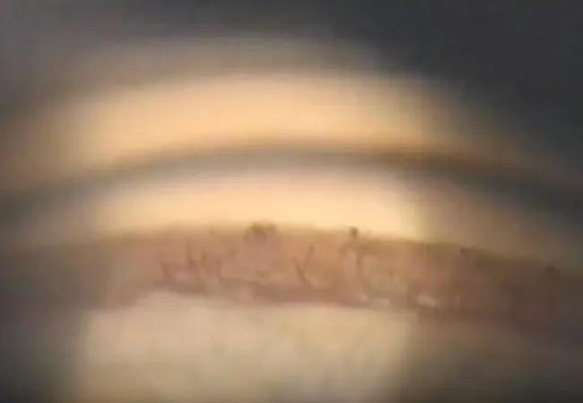
Image content: This image is available to view online.
View image online (https://assets.clevelandclinic.org/transform/b5858bee-3303-4e78-bf82-906844203451/neovascular-glaucoma)
Neovascular glaucoma
Pictured above: Gonioscopy image of neovascular glaucoma
Advertisement
Cleveland Clinic is a non-profit academic medical center. Advertising on our site helps support our mission. We do not endorse non-Cleveland Clinic products or services. Policy
By Mary Qiu, MD, and Jessie Wang, MD
Neovascular glaucoma (NVG) is an aggressive form of secondary glaucoma resulting from retinal ischemia caused by conditions like diabetic retinopathy, retinal vein occlusion and ocular ischemic syndrome. Initially the angle is open despite neovascularization, but the disease progresses to a closed-angle glaucoma with the formation of peripheral anterior synechiae (PAS).
Historically treatment for NVG has been guided by presumed visual potential. Retina specialists can perform panretinal photocoagulation (PRP) to suppress the neovascular drive, while glaucoma specialists can use tube shunts or diode laser technology to lower intraocular pressure (IOP).
However, even when patients with NVG get appropriate treatment, the outcomes are often poor. A 2021 study reported that 15% of patients with NVG treated with tube shunts still developed no-light-perception blindness, which may be because placing a tube doesn’t control the retinal etiology of the disease.
Now, with the rise of anti-VEGF injections and microinvasive glaucoma surgery, we have more options. Angle surgeries can lower IOP by preserving the conventional outflow pathway. This new era of treatment for both retina disease and glaucoma offers new hope for better outcomes in patients with NVG.
The best treatment for NVG depends on the stage of disease, including:
Advertisement
Treating patients as early as possible, while accounting for angle anatomy, leads to better outcomes. As such, a gonioscopy is recommended as part of the initial assessment for every NVG patient.
IOP-lowering medications either decrease the amount of fluid the eye makes or increase the amount of fluid the eye drains. However, the drain only works if it is not blocked by scar tissue.
What happens when medication doesn’t lower IOP? Many providers would insert an Ahmed tube shunt, which would lower the IOP right away, but performing this surgery in the acute stage has a higher risk of bleeding-associated complications.
Instead, for eyes with NVG with 360 degrees of PAS and active neovascularization, we recommend promptly performing nonincisional primary cyclophotocoagulation (CPC). This decreases the amount of fluid the eye makes without going into the eye or causing bleeding. Then, you can follow up with anti-VEGF injection and other treatments once the condition is stabilized.
What’s important to note is that CPC is not only for eyes with poor visual potential. When a patient presents with acute NVG, we may not know what their actual visual potential is.
To control the underlying retinal disease, a retina specialist can provide anti-VEGF injections and PRP to decrease the neovascular drive. PRP gradually reduces neovascularization or prevents it. It does not act quickly, however, so it isn’t the best for emergent conditions.
Anti-VEGF injection can have the same effect as PRP, but it works much quicker, sometimes within one day. This is helpful to temporarily regress vessels, so more steps can be taken to reduce the IOP as soon as possible. More treatment can be done later to permanently regress the vessels.
Advertisement
If the angle is open or partially open, anti-VEGF may help lower the IOP as well. It will not lower IOP if the angle is closed and the drain is blocked. Again, we can predict how a patient’s eye will respond by performing gonioscopy.
When first examining a patient with NVG, performing a gonioscopy is the most important thing you can do to help determine next steps in treatment. Treatment for an eye with an open angle is completely different from treatment for an eye with a closed angle.
In addition, sustained collaboration with retina colleagues is essential. NVG is a complex, multidisciplinary condition, and good outcomes depend on personalized decision-making and long-term control of the retinal disease.
Drs. Qiu and Wang are glaucoma specialists at Cleveland Clinic Cole Eye Institute. This article was based on their booth presentation at the 2025 American Academy of Ophthalmology meeting.
Advertisement
Advertisement

Prescribing eye drops is complicated by unknown risk of fetotoxicity and lack of clinical evidence

A primer on MIGS methods and devices

7 keys to success for comprehensive ophthalmologists

From medication to laser treatment to surgery

Tissue remnants seem unrelated to clinical outcome
Minimally invasive surgery is effective for uveitic and steroid-induced glaucoma too

The drug does not provide lasting disease control for most patients

New service for patients with diabetes will make annual screening more convenient