Locations:

A hand and wrist surgeon explains different approaches based on nature and severity of injury
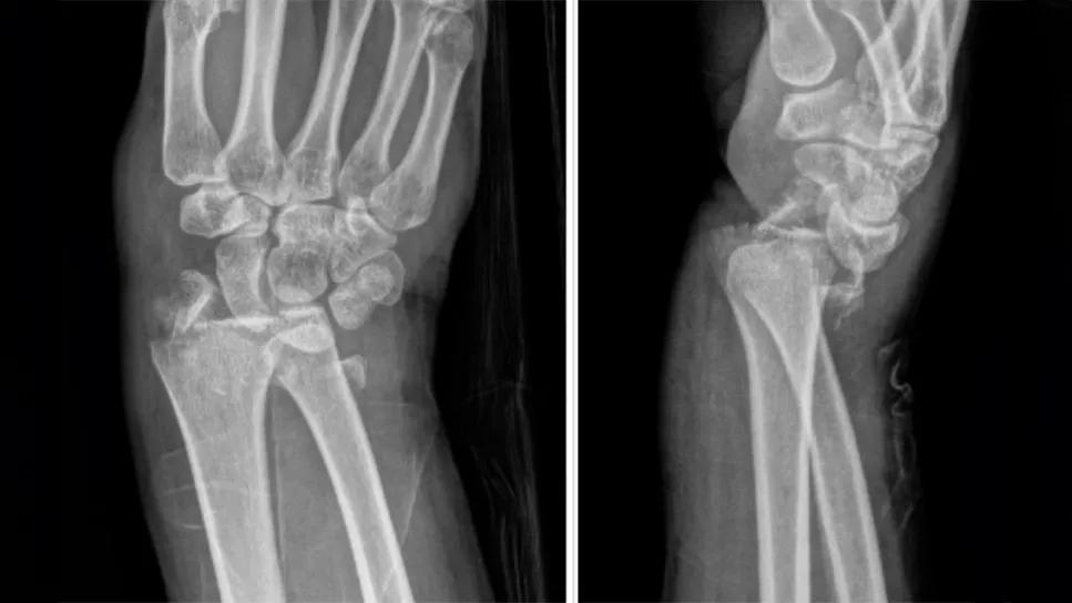
Image content: This image is available to view online.
View image online (https://assets.clevelandclinic.org/transform/e7800d1a-c02f-4816-9d8b-b71fccc22eec/comminuted-intra-articular-distal-radius-fractures-feature)
X-ray of a broken wrist
Advertisement
Cleveland Clinic is a non-profit academic medical center. Advertising on our site helps support our mission. We do not endorse non-Cleveland Clinic products or services. Policy
Distal radius fractures remain one of the most commonly encountered musculoskeletal injuries, representing a spectrum of pathologies from simple extra-articular fractures to complex intra-articular and comminuted fractures.
Hand and wrist surgeons recognize that the decision-making process for surgical intervention must be nuanced, reflecting not only the fracture pattern but also the patient’s expectations, bone quality and functional demands.
Here, we discuss the different fracture patterns, surgical indications and variations in surgical strategies for distal radius fractures, with a focus on advanced treatment modalities and the importance of tailoring management to the specific characteristics of the patient and injury.
Extra-articular fractures are among the most common distal radius fractures. They do not involve the articular surface of the radiocarpal joint. Most often they are caused by low-energy falls, such as falls from standing in patients with osteopenic or osteoporotic bone. These injuries are consistent with fragility fractures. Extra-articular fractures also can occur in a younger patient population, generally from high-energy mechanisms, such as falls from a height (e.g., ladder), sports-related injuries and motor vehicle accidents.

Image content: This image is available to view online.
View image online (https://assets.clevelandclinic.org/transform/623a4ead-c600-4181-ac0e-1ee03e4da4c5/radiocarpal-fracture-dislocations)
Extra-articular fracture
While often less complex than intra-articular fractures, these injuries still require careful assessment. Nonoperative treatment is effective for stable, nondisplaced fractures, while surgical intervention (e.g., open reduction and internal fixation or external fixation) is necessary for displaced, unstable or comminuted fractures.
Advertisement
Surgeons must consider the fracture’s characteristics as well as the patient’s age, bone quality and functional demands to determine the most appropriate treatment. With early and appropriate intervention, most patients with extra-articular distal radius fractures can expect a good functional outcome.
Radiocarpal fracture-dislocations are complex and potentially devastating injuries that involve both a fracture and dislocation of the radiocarpal joint. These injuries are often the result of high-energy trauma, such as motor vehicle accidents or falls from height.
They may be associated with significant soft tissue injuries (including soft tissue and ligament disruption) as well as neurovascular injury. Early diagnosis and appropriate management are crucial to achieving optimal functional outcomes and minimizing long-term complications such as arthritis, chronic instability and compromised wrist function.

Image content: This image is available to view online.
View image online (https://assets.clevelandclinic.org/transform/81016933-6d09-47d0-ba25-c93e5525b48b/fractures-with-volar-marginal-fragment)
Radiocarpal fracture-dislocation
Initial management includes a thorough clinical exam, assessment of the soft tissue envelope and detailed neurovascular exam. Fracture reduction and manipulation should be attempted if the fracture is reducible, particularly to offload the soft tissue envelope while definitive management is planned.
Given the unstable nature of these injuries, surgical fixation is typically indicated to restore stable anatomic alignment. This may involve the use of volar locking plates, dorsal spanning plate fixation versus external fixation, or fragment-specific fixation depending on the fracture pattern, associated ligamentous injuries and bone quality.
Advertisement
Postoperatively, the wrist is generally immobilized. The duration of immobilization depends on the fracture complexity, fixation construct and patient factors. Postoperative rehabilitation is critical to restoring function and minimizing complications.
Intra-articular distal radius fractures are complex injuries involving the articular surface of the distal radius. They often lead to joint instability, functional impairment and long-term complications like arthritis.
A subset of these fractures involves a volar marginal fragment, a distinct pattern of intra-articular distal radius fractures that requires careful attention. This type of fracture is particularly challenging as it is often intraoperatively missed due to its small size and volar extrinsic ligament attachments. Failing to capture this fragment with internal fixation creates the potential for joint incongruity and persistent instability.

Image content: This image is available to view online.
View image online (https://assets.clevelandclinic.org/transform/5ff0c588-52d4-4b65-af09-ef42388a82ee/comminuted-intra-articular-distal-radius-fractures)
Intra-articular fracture with volar marginal fragment
In addition to X-ray imaging, preoperatively these fractures often benefit from CT imaging to better visualize the fracture configuration, particularly the volar marginal fragment. CT imaging also is useful in determining the degree of comminution and the potential need for fragment-specific fixation.
Given the inherently unstable nature of these injuries, surgical fixation is generally recommended. I perform an extended flexor carpi radialis approach for these fractures to achieve adequate visualization of the central column and the volar marginal fragment, which aids in achieving an accurate reduction of the fracture. Fixation of this fracture pattern often entails use of a volar locking plate with or without additional miniplates, screws or K-wires to stabilize the volar marginal fragment while restoring articular congruency.
Advertisement
Postoperatively, we follow these cases more closely with X-ray imaging and clinical exam to confirm maintenance of fracture reduction and appropriate progress with range of motion.
Comminuted intra-articular distal radius fractures are among the most challenging injuries encountered in orthopaedic practice. These fractures typically result either from high-energy trauma, such as falls from significant heights or motor vehicle accidents, or from low-energy falls in patients with osteopenic or osteoporotic bone.
These fracture patterns involve significant disruption of the distal radius, with multiple fracture fragments that extend into the radiocarpal joint, compromising the articular surface. The complexity of the injury often leads to poor functional outcomes if not properly managed.
Early recognition, precise fracture reduction and stable fixation are critical for optimizing wrist function and preventing long-term complications, such as post-traumatic arthritis and joint instability.
In addition to X-ray imaging at the time of injury, a CT scan is often necessary for comminuted intra-articular fractures. It allows for more detailed visualization of the fracture fragments, especially when there is concern about the exact alignment of the articular surface and the degree of comminution. A CT scan also is helpful for selecting the type of fixation.

Image content: This image is available to view online.
View image online (https://assets.clevelandclinic.org/transform/85cd8658-5ae2-48fe-a90d-4c033b85de7f/associated-carpal-tunnel-syndrome)
Comminuted intra-articular fracture
Surgical treatment is often recommended for these fractures given their poor prognosis with nonoperative management. Depending on the fracture pattern, fixation can be achieved with a volar approach, a dorsal approach or arthroscopic-assisted fixation. I prefer a volar approach coupled with dry arthroscopic-assisted fixation. This approach significantly improves the accuracy of recreation of the articular surface while allowing for full assessment of any associated soft tissue injury, such as tears in the scapholunate ligament or triangular fibrocartilage complex.
Advertisement
For all distal radius fractures except the most common extra-articular fractures, I assess for carpal tunnel symptoms preoperatively.
Carpal tunnel syndrome can occur particularly in fractures that result from high-energy mechanisms or in the setting of significant fracture displacement. Patients generally present with paresthesias in the median nerve distribution (thumb, index, middle and radial aspect of ring finger) arising after injury.
If I suspect the presence of carpal tunnel syndrome, a key decision revolves around whether to manage it conservatively or proceed with surgery. This decision is influenced by acuity and persistence of symptoms despite an appropriate reduction, edema control and splint management.
In patients with a distal radius fracture and concurrent carpal tunnel syndrome that persists despite conservative measures, I prefer to perform an endoscopic carpal tunnel release at the time of surgical fixation of their fracture. The advantages of an endoscopic carpal tunnel release include reduced soft tissue dissection and, therefore, reduced morbidity of surgery in a setting that requires open surgery for fracture fixation. This minimally invasive approach (involving a 1 cm incision ulnar to the distal radius exposure) avoids a palmar incision that could increase the risk of postop pain and lead to delayed rehab. It is always possible that an endoscopic carpal tunnel release would need to be converted to an open procedure particularly in the trauma setting.
Distal radius fractures are among the most common fractures, and treatment often requires balancing functional recovery, preventing complications and minimizing the risk of long-term disability.
The nature and severity of the fracture is paramount in determining whether surgery is needed. However, the decision to proceed with surgery should always consider the patient’s functional demands, comorbid conditions and expectations.
Dr. Marjoua is staff in the Department of Orthopaedic Surgery at Cleveland Clinic. She specializes in hand and wrist surgery.
Advertisement
Trauma patients present with infected pilon and distal femur fractures
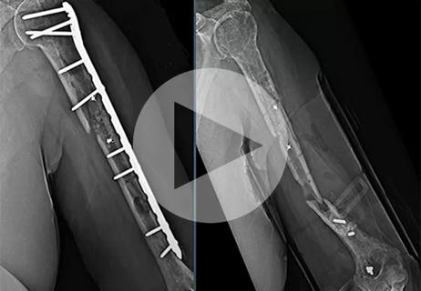
Finally, a solution after multiple revision surgeries for delayed bone healing, loose hardware and unrelenting infection
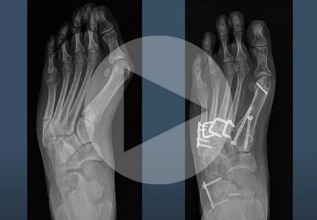
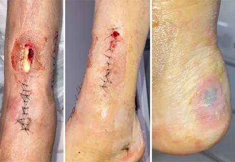
Surgeon corrects skew foot to address repeat injuries

Even patients with reported penicillin allergies can receive it without increased complications

Study challenges assumptions about risk evaluation in total hip revision

Protein expression in synovial fluid indicates patients’ immune factors may be involved

Cleveland Clinic’s Global Peak Performance Center and PGA TOUR partnership pair advanced assessment with longitudinal follow-up to enhance clinical decision-making

Study highlights the need for objective functional measures as value-based care expands