Locations:

Multimodal evaluations reveal more anatomic details to inform treatment
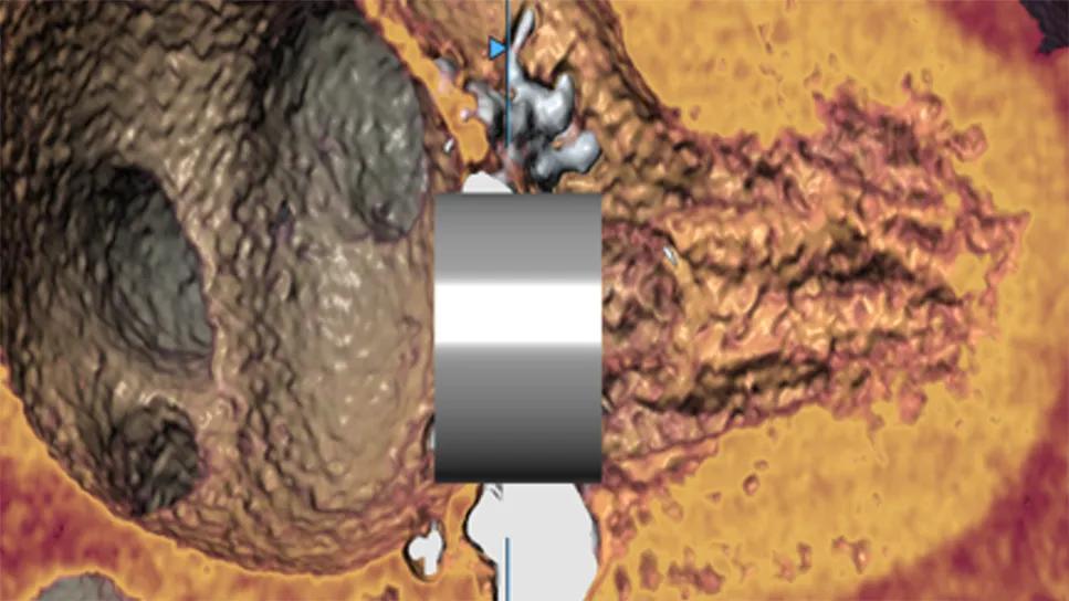
Image content: This image is available to view online.
View image online (https://assets.clevelandclinic.org/transform/d1c74820-009e-4d5c-a406-e6e477530083/valve-simulation-cqd)
3D heart imaging study with metal cylinder overlay
When a patient presents with symptoms of mitral valve disease, listening to the patient and listening to their heart may begin to reveal a diagnosis. However, imaging tells a fuller story.
Advertisement
Cleveland Clinic is a non-profit academic medical center. Advertising on our site helps support our mission. We do not endorse non-Cleveland Clinic products or services. Policy
“Imaging captures everything we need to know about a patient’s mitral valve disease,” says Brian Griffin, MD, Section Head of Cardiovascular Imaging at Cleveland Clinic and Medical Director of its Valve Center. “Is it stenosis, regurgitation or a combination? What’s the underlying cause? How severe is it in terms of hemodynamics? What is the impact on heart function? What’s the best way to manage the condition?”
Cleveland Clinic’s cardiovascular imaging physicians and technologists are experts at selecting, conducting and coordinating a multitude of imaging modalities, from the trusty workhorse echocardiography (including transesophageal, intraoperative, intracardiac and stress echo) to more specialized technologies such as MRI, CT and nuclear scans.
“Our ability to masterfully conduct and interpret all of these modalities ensures that we get the maximum information about a patient’s condition,” Dr. Griffin says. “Most of our cardiovascular imaging physicians have expertise in multiple modalities, so after they analyze one set of scans they can identify the best imaging technology to answer any remaining questions about a case. Since each modality provides different details, many times we coordinate tests to perform a true multimodal imaging evaluation.”
In this article, imaging experts discuss the latest advances in three imaging modalities that Cleveland Clinic often uses when assessing and managing mitral valve disease.
Transthoracic echocardiography is the cornerstone of mitral valve evaluation, says Cleveland Clinic cardiovascular imaging specialist Rhonda Miyasaka, MD. It’s the starting point of any mitral valve case and easily repeated to assess changes over time.
Advertisement
“In the past few years, we’ve had a lot of advancement in image quality as well as 3D technology, which many people associate only with transesophageal echo,” Dr. Miyasaka says. “At Cleveland Clinic, we apply 3D imaging techniques to transthoracic echo as well and can get amazing images of the mitral valve. I tell people not to underestimate the power of echo.”
Transesophageal echo (TEE) continues to supplement transthoracic evaluations with closer views and more detailed images of mitral valve anatomy, which can help isolate leaks, reveal why leaks are occurring and inform treatment.
“These details are clinically relevant because of the broad range of treatment options now available,” Dr. Miyasaka says. “By viewing the leaflet morphology, chordae tendineae, clefts and other anatomic details, we are better able to determine if the patient should be recommended for open surgery, transcatheter intervention or something else.”
Advances in 3D imaging also have benefited transcatheter interventions, which rely on TEE for intraprocedural guidance.
“TEE techniques are going to get better and better, producing even higher quality images, which can open the door for new devices and treatment technologies,” Dr. Miyasaka predicts.
CT imaging provides excellent anatomic detail with high spatial resolution. Calcifications are readily identified on CT, and in situations where ultrasound is limited by acoustic shadowing from prosthetic materials such as rings and valves, CT can be particularly valuable, explains Cleveland Clinic cardiovascular imaging specialist Serge Harb, MD.
Advertisement
CT imaging also plays an important role in planning:
“CT is also crucial when planning transcatheter mitral valve replacement,” Dr. Harb says (see Figure 1). “It allows you to accurately measure the annulus, virtually embed a simulated valve to evaluate how it will sit, and anticipate any potential complications.”

Image content: This image is available to view online.
View image online (https://assets.clevelandclinic.org/transform/ada6ce4f-8899-40a0-8f2f-a105e1bd7f46/advanced-simulation-cqd-image1)
Figure 1. The utility of CT imaging in planning transcatheter valve replacement in severe mitral annular calcification. CT helps assess the distribution of annular calcification (top left), simulate valve replacement (top middle), and predict risks such as paravalvular leak (top right) and left ventricular outflow tract obstruction (LVOTO) based on neo-LVOT measurement (bottom).
Dr. Harb predicts that the utility of CT imaging will expand as minimally invasive and transcatheter mitral valve procedures evolve.
“When working through a small incision or catheter without full exposure to the mitral valve, CT imaging provides the preprocedural visualization needed to ensure a safe and effective intervention,” he says.
When there is uncertainty about severity of mitral valve disease or a discrepancy between a patient’s symptoms and what is seen on echo or CT, cardiac MRI (CMR) can add quantifiable detail (Figures 2 and 3). It is the gold standard for quantifying ventricular and atrial size and function in patients with mitral regurgitation, says Deborah Kwon, MD, Director of Cardiac MRI at Cleveland Clinic. It also can reliably reveal and quantify the extent of ventricular fibrosis (and possible associated risk of arrhythmia), not to mention underlying myocardial or ischemic injury that may be contributing to mitral valve dysfunction.
Advertisement

Image content: This image is available to view online.
View image online (https://assets.clevelandclinic.org/transform/26101707-b8cd-42dd-869d-7d96fe6d906e/advanced-simulation-cqd-image2b)
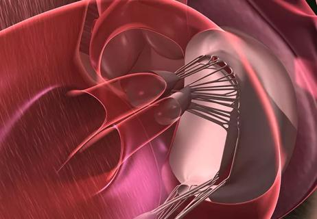
Figure 2. The utility of cardiac MRI in assessing secondary mitral regurgitation.

Image content: This image is available to view online.
View image online (https://assets.clevelandclinic.org/transform/89f94192-3ba8-48d4-b161-267dd9f4ec2d/advanced-simulation-cqd-image3)
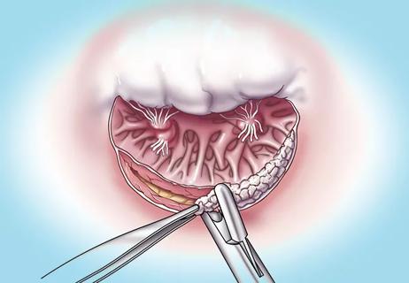
Figure 3. The utility of cardiac MRI in assessing primary mitral regurgitation.
“MRI is the most comprehensive imaging modality not only for looking at mitral valve dysfunction but also for assessing its causes and effects in the unique context of underlying myocardial tissue characteristics,” Dr. Kwon says.
Cardiac MRI remains less widely available in the U.S., largely due to the challenges associated with acquiring CMR images and the limited number of skilled cardiac MRI technicians. To address this, the team at Cleveland Clinic is working to expand access by developing a CMR technique that enables any hospital with an MRI magnet, even centers that don’t have a dedicated cardiac MRI technician, to acquire cardiac MRI scans.
“We’re still in the research phase, but it is exciting that other solutions with innovative artificial intelligence-based technologies have been emerging that likely will collectively and broadly expand CMR access,” Dr. Kwon says. “Cardiac MRI used to be a niche specialty available only at certain centers, but utilization has been steadily increasing as multiple studies are demonstrating its impact on risk prognostication. Consequently, international guidelines now recognize cardiac MRI with Class 1 indications for assessment of valvular heart disease for certain indications. Additionally, a statement recently published by the American College of Cardiology and the American Heart Association recommends Level 1 CMR training as part of general cardiology curriculum, alongside core competencies for echocardiography, nuclear imaging and CT. Therefore, we in the field need to standardize and democratize cardiac MRI access and interpretation so more patients can benefit from it.”
Advertisement
Cleveland Clinic is fortunate to have multiple imaging technologies that help identify different aspects of mitral valve conditions and present a complete picture, says Dr. Griffin.
“We are always seeking more information and researching new protocols for identifying abnormalities in tissue,” he says. “We have published extensively on indices of mitral valve disease (J Am Coll Cardiol. 2016;68[18]:1974-1986), particularly on the timing of intervention. When you have mitral valve malfunction, damage can be occurring to heart function on a subclinical level. In general, if the mitral valve is leaking badly and the valve appears to be repairable, then the sooner we address the mitral valve, the better — even if the patient is asymptomatic. Imaging will continue to teach us more about the best timing of treatment.”
Advertisement

Scenarios where experience-based management nuance can matter most

Optimal management requires an experienced center

Provides option for patients previously deemed anatomically unsuitable
While mortality was unaffected, later surgery was associated with more reoperations
Surgical tips on debridement of calcification to optimize valve replacement
Superiority continues even after significant crossovers from control group at 2 years

Join us in New York Dec. 4-5 for evidence-based instruction with real-world examples

CMR-CLIP outperforms general AI tools; may one day expand patient access to CMR