Locations:

A new online calculator can determine probability of melanoma
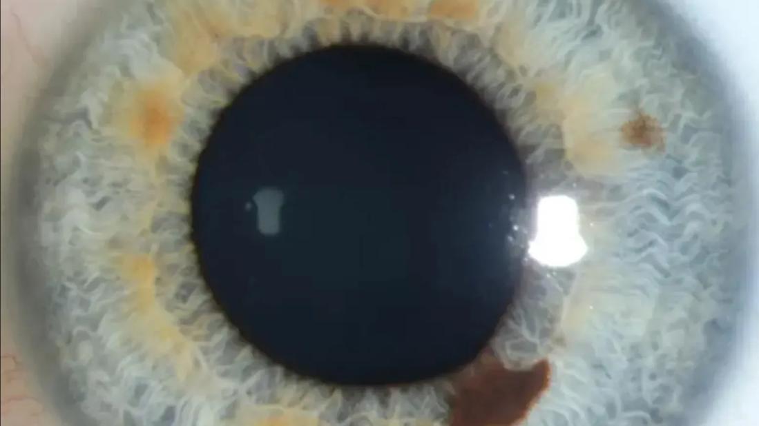
Image content: This image is available to view online.
View image online (https://assets.clevelandclinic.org/transform/3f7ea99f-d1c5-4432-ac44-91d5ad6f3cdd/iris-freckle)
Eye with a lesion on the iris
Iris melanoma may be the least common type of uveal melanoma, but it can be the most conspicuous. A lesion in the colored part of the eye, visible under the transparent cornea, is hard to miss. But it’s easy to confuse iris melanoma with benign growths, such as freckles and nevi.
Advertisement
Cleveland Clinic is a non-profit academic medical center. Advertising on our site helps support our mission. We do not endorse non-Cleveland Clinic products or services. Policy
“Melanoma of the iris is very rare, affecting about one in 1 million people,” says Arun D. Singh, MD, Director of Ophthalmic Oncology at Cleveland Clinic Cole Eye Institute. “I’ve seen it mostly in people with blue, light brown or other light irises — mostly people with Northern European ancestry. We don’t have strong data that links the melanoma to sun exposure or any occupation.”
Dr. Singh explained more about the diagnosis and treatment of this rare eye cancer in a recent episode of Cleveland Clinic’s Cancer Advances podcast.
Iris melanoma is usually detected early, when it’s small.
“Everybody looks at their own eyes many times a day, so a lesion is often noticed quickly,” Dr. Singh says. “It’s common to diagnose an iris melanoma when it’s only 3-4 mm — very small compared to melanoma elsewhere on the body. Catching it while it’s still small means there’s a lower risk of metastasis.”
Surgical excision is the standard treatment. The iris will not regenerate, but usually surgeons can pull and stitch remaining areas of the iris to recreate a pupil. If the defect is large, the pupil may not be able to be recreated. Radiation therapy is an effective alternative to surgery.
“Outcomes are excellent,” Dr. Singh says. “We estimate that the risk of metastasis is only about 1% over five years. Local recurrence is 1% to 5% — closer to 1% after surgery or radiation. Treatment focuses on local control, and if you can achieve local control of iris melanoma, it’s like achieving a cure.”
When a spot appears on a patient’s iris, the first step for any physician is to photograph it.
Advertisement
“If it’s small, it’s more likely something benign, like a nevus or freckle,” Dr. Singh says. “Those things are much more common than iris melanoma. It can be hard to tell the difference between those lesions, so it’s important to see if the spot grows or changes over time. A baseline photograph can help.”
A spot that has been there for years and not changed is likely to be benign. A spot that changes or appears suddenly may require more evaluation.
Diagnosing iris lesions of any type is a clinical task, done without surgical biopsy or other invasive techniques.
Iris freckles tend to look like tiny, smudge-like spots, Dr. Singh notes. Iris nevi are more confluent and solid-looking.
According to Dr. Singh’s 2024 study published in the British Journal of Ophthalmology, iris freckles have a median largest basal diameter of 0.8 mm (compared to 2.1 mm for nevi) and a median thickness of 0.04 mm (compared to 1.0 mm for nevi). Freckles tend to appear in groups and are located centrally. Unlike iris nevi, iris freckles have irregular margins and appear flat on anterior segment optical coherence tomography (AS-OCT). They are not detectable by ultrasound biomicroscopy (UBM).
Melanoma lesions can be identified by clinical characteristics, including:
Melanoma is suspected if the lesion:
Advertisement
In a second study recently published in the British Journal of Ophthalmology, Dr. Singh and Emily Zabor, DrPH, introduced a prediction model for iris melanoma. Developed by evaluating 97 cases of iris melanoma and 112 cases of iris nevus seen at the Cole Eye Institute, the model calculates the probability of melanoma according to a lesion’s basal diameter, height, location and other features. Physicians can use this calculator to inform decision-making about treating or referring patients with iris lesions.
This Iris Melanoma Predictor is similar to another online tool introduced in 2021 to predict choroidal melanoma.
“Currently, someone needs to review images of a lesion and manually enter data into the model,” Dr. Singh says. “Our goal is to automate the process, having AI analyze the image and predict a diagnosis.”
In addition to using this prediction model, physicians should consider referring any patient with a new or concerning iris lesion to a specialized center for evaluation, he concludes.
Advertisement
Advertisement

The drug does not provide lasting disease control for most patients

A review of pathophysiology, risk factors, clinical presentation and treatment options

Registry data highlight visual gains in patients with legal blindness

Study is first to show reduction in autoimmune disease with the common diabetes and obesity drugs

It’s the first step toward reliable screening with your smartphone

Fixational eye movement is similar in left and right eyes of people with normal vision

Oral medication may have potential to preserve vision and shrink tumors prior to surgery or radiation
Only 33% of patients have long-term improvement after treatment