Locations:

Treatment insights from the ninth recorded EMPD case in 50 years
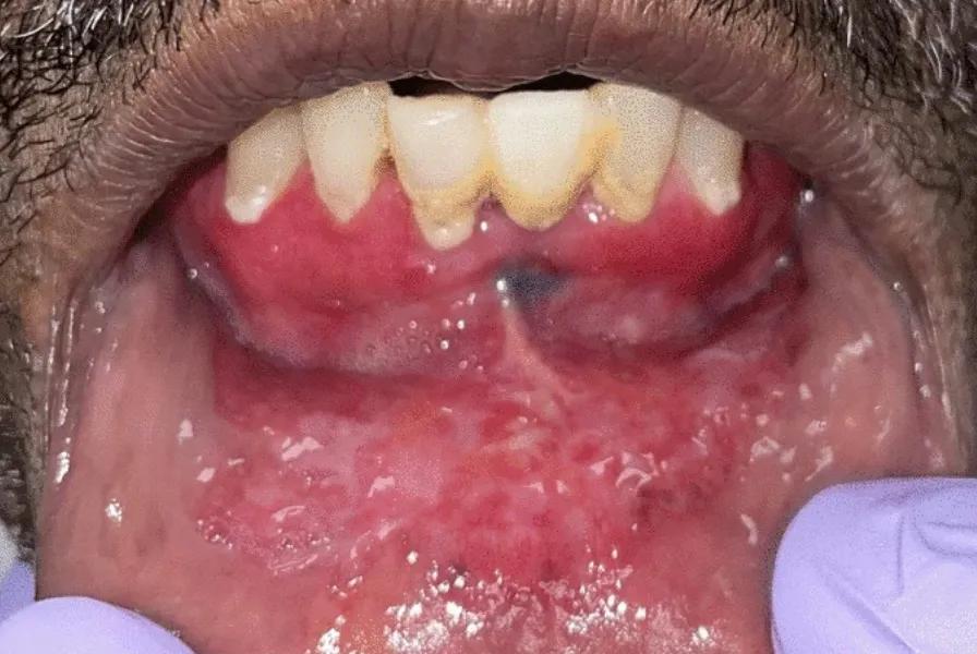
Image content: This image is available to view online.
View image online (https://assets.clevelandclinic.org/transform/15b0fb09-8180-4e8a-8843-a354a7e07c12/Extramammary-PagetDisease-OralMucosa-figure1A)
View of lower lip showing EMPD
A 72-year-old man with a history of smoking presented with a diagnosis of Extramammary Paget disease of his oral mucosa. The patient had been seen by a community-based oral and maxillofacial surgeon due to unexplained weight loss over one year, as well as tenderness of his oral mucosa when eating, drinking and toothbrushing over the previous two months. An examination of the patient’s mouth revealed what appeared to be irritation of the mucosal lining.
Advertisement
Cleveland Clinic is a non-profit academic medical center. Advertising on our site helps support our mission. We do not endorse non-Cleveland Clinic products or services. Policy
The surgeon initially suspected lichen planus and referred the patient for a biopsy. However, a biopsy confirmed a diagnosis of Extramammary Paget disease (EMPD), and the patient was referred to Cleveland Clinic head and neck surgeon Danielle Bottalico, MD, for a surgical consult.
Typically, this condition is seen in breast carcinoma, where it is diagnosed as mammary Padget disease (MPD). Very few cases have been seen outside of breast tissue.
“Extramammary Paget disease is exceptionally rare,” Dr. Bottalico explains. “To the best of our knowledge, it’s only been documented eight times, and ours was the ninth case report over the past 50 years.”
Because EMPD is so rare, its risk factors and causes are not known. However, it appears to mimic an inflammatory process and can be associated with underlying invasive adenocarcinoma.
In this specific case, a CT scan was performed, and no underlying mass concerning for cancer was identified.

Image content: This image is available to view online.
View image online (https://assets.clevelandclinic.org/transform/5b01d79f-4eb4-4be4-a17c-104f58930cda/Paget-Cell-Micrograph)
Above: Paget cells exhibit markedly atypical nuclei with abundant amphophilic cytoplasm. Top of page: Extensive red/white mucosal change of anterior mandibular vestibule and lower lip concerning for lichenoid inflammatory process or dysplasia/carcinoma. Both images reprinted from Dababneh et al, Head Neck Pathol. 2024;18[1]:33, under the Creative Commons CC BY license. © The Authors
The case was presented to Cleveland Clinic’s multidisciplinary tumor board, which includes a panel of head and neck surgical oncologists, radiologists, radiation oncologists, medical oncologists and pathologists to consult on the appropriate treatment.
Management for oral EMPD is primarily surgical, and given the extent of involvement the case was also referred to Cleveland Clinic head and neck surgeon Jamie Ku, MD, for reconstruction.
For the surgery, Dr. Bottalico performed a wide local excision, removing all involved mucosa and a marginal mandibulectomy. Head and neck pathologist Ivan Stojanov, DMD, examined the specimen while the patient was still asleep, to determine if the margins were clear.
Advertisement
“Some of the tissue that had initially appeared normal was actually affected by the disease, and I had to go back and re-resect the area of concern until Dr. Stojanov and his fellow could confirm there was no microscopic disease,” Dr. Bottalico notes.
Dr. Ku performed the reconstruction, using tissue from the patient’s forearm, and the reconstructive surgery went as planned.
The patient’s recovery went smoothly, and he was discharged about one week after surgery. He did not receive any adjuvant therapy and remains free of disease approximately 18 months after surgery.
Because EMPD can recur, the patient comes in for regular follow-up visits. Postoperative monitoring has included CT scans with contrast, with baseline imaging performed after surgery, and follow-up scans conducted at six months and one year. The patient will continue to be followed for surveillance.
Dr. Bottalico notes that expected outcomes for EMPD depend on whether the disease is associated with an underlying cancer.
“This patient had no underlying malignancy, and as a result, I think his prognosis is quite good,” notes Dr. Bottalico.
Although the causes of this rare disease are unknown, it has been hypothesized that EMPD, as well as rare cases of MPD that are not associated with underlying breast cancer, may originate from Toker cells. These are CK7 immunopositive cells that are morphologically similar to Paget cells and are thought to be potential precursor cells to Paget disease.
In this case, Dr. Bottalico notes that cells from the patient’s specimen also stained positive for CK7.
Advertisement
Providers should know that, although rare, EMPD can masquerade as lichen planus or dysplasia. Biopsies should be examined by pathologists who are familiar with this condition.
For patients diagnosed with EMPD, it’s important to know that the disease can be associated with underlying malignancies.
“Because this is very rare, this patient’s work up and treatment involved expertise from our multidisciplinary head and neck team here at Cleveland Clinic,” Dr. Bottalico says.
The team’s paper, “Extramammary Padget Disease of Oral Mucosa: Case Report With Literature Review,” was published in Head and Neck Pathology.
Advertisement
Advertisement

Scleromyxedema may cause extracutaneous symptoms

Treatment strategies require understanding of pathomechanisms

Major study demonstrates importance of having a multidisciplinary approach to treatment for large, locally advanced tumors

How effective management integrates multiple specialties and therapies

Systematic review indicates more can be done for higher-risk patients

Analysis of HNSCC patients shows HPV status to be predictive of higher abundance of oncobacteria within the tumor

Case study illustrates the potential of a dual-subspecialist approach

Evidence-based recommendations for balancing cancer control with quality of life