Locations:

Challenging case requires outside-the-box approach
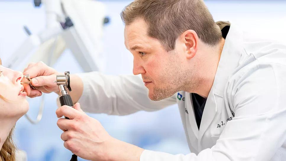
Image content: This image is available to view online.
View image online (https://assets.clevelandclinic.org/transform/5aa0d04d-c51d-43de-a6f4-32731bd6f4c4/23-HNI-4354408-NasalSeptalPerforation-CaseStudy-CQD_967x544-1_jpg)
Dane Genther, MD
Nasal septal perforation — a full-thickness defect of the nasal septum — occurs for several reasons and can have a significant impact on quality of life. Patients with this condition may experience a variety of issues including whistling, crusting, nasal obstruction/congestion and nasal dyspnea, among others.
Advertisement
Cleveland Clinic is a non-profit academic medical center. Advertising on our site helps support our mission. We do not endorse non-Cleveland Clinic products or services. Policy
While some patients can be managed with more conservative measures, such as nasal emollients and rinses, other cases may require surgical intervention. Nasal septal perforation repair is a complex procedure, and there are a variety of techniques that a surgeon may choose.
Recently, Dane Genther, MD, Facial Plastic and Reconstructive Surgeon, Otolaryngology-Head and Neck Surgery at Cleveland Clinic, successfully performed a nasal septal perforation repair using a novel approach, offering another potential treatment option for these patients.
A female patient was diagnosed with a nasal septal perforation approximately two years ago. “This condition changes the way airflow feels in your nose, and so, for many people, it is very anxiety-provoking,” notes Dr. Genther. “In this case, the patient was experiencing significant nasal drainage and underwent two repairs at other facilities, both of which failed.”
The first repair used a hybrid method that involved a local tissue rearrangement flap and some temporalis fascia from the scalp, explains Dr. Genther. The second surgery was a similar, but more extensive procedure that used the other side of the temporalis fascia scalp.
After the second failed attempt, the patient came to Cleveland Clinic where Dr. Genther took over her care with support from his team, which included resident Stephen Hadford, MD. While he typically would use a temporalis fascia in this case, both areas where he would have harvested fascia were used in the previous surgeries. Therefore, a modified approach was required.
Advertisement
“The mechanism behind this technique was similar to what we normally do with the temporalis fascia, but instead we used fascia lata from the thigh, which is a thicker piece of fascia,” Dr. Genther explains. “Fascia lata is very strong and has a variety of uses, and the reason that it works is because it has this areolar layer of small blood vessels around it, which likely allow it to be revascularized fairly quickly and heal. When we put it between the two sides of the perforation it acts as a scaffold for the mucosa to grow over and the two sides heal separately so there’s not a perforation.”
Like their usual technique, which Dr. Genther and colleagues outlined in a recent paper (Am J Otolaryngol 2023;44(4):103883), a polydioxanone (PDS) absorbable plate is used to stabilize the graph. “This stabilizes it more rigidly temporarily until it dissolves,” he explains. “In this patient’s case, we then used the fascia lata instead of the trilayer temporalis fascia.”
When discussing the challenges of this particular case, Dr. Genther notes, “Since there had already been two attempts to repair the perforation, it was a difficult and slow process to open everything up and keep the flaps intact because the last thing you want to do is create more holes. So, it took a long time because there was previous graft material and significant scar tissue.
“However, the rest of the procedure was fairly straightforward and routine,” he adds. “The key is being extra careful with the fascia and leaving that vascular layer on the outside. With every surgery I perform, I get more careful with that vascular layer, and I think that is where other people using similar techniques come up short.”
Advertisement
Following the procedure, the splints were left in for one month. Dr. Genther has since seen the patient for two follow-up appointments and reports that her perforation is healed. “Everything essentially looks normal on the inside,” he says. “She’s breathing better while still having some drainage; however, that is expected. Overall, she is doing great and healing well. We will follow up again in about six months.”
While Dr. Genther has been interested in this approach, this is the first time he has used it in clinical practice. Based on the success of this procedure, he plans to continue to explore this technique at Cleveland Clinic and its impact on patients with nasal septal perforation.
“Our team at Cleveland Clinic has extensive experience with newer techniques, which allows us to not only manage these complex patients, but also ensure positive outcomes for even the most challenging cases,” Dr. Genther says, while emphasizing that the case described above demonstrates that this approach works, and he looks forward to offering it to more patients. “This technique gives surgeons a new way to successfully repair nasal septal perforation and improve outcomes and quality of life for their patients.”
Podcast content: This podcast is available to listen to online.
Listen to podcast online (https://www.buzzsprout.com/2241209/16533037)
Advertisement
Advertisement
Collaboration critical to a successful resection

Study provides Class III evidence that assay distinguishes disease from normal controls

Surprising X-ray results explain a snoring sound heard during breathing
The disorder can greatly impact patients’ quality of life, but sclerotherapy and a multidisciplinary approach to care can be life-changing

How effective management integrates multiple specialties and therapies

Systematic review indicates more can be done for higher-risk patients

Analysis of HNSCC patients shows HPV status to be predictive of higher abundance of oncobacteria within the tumor

Case study illustrates the potential of a dual-subspecialist approach