Locations:


First reported intestinal autotransplantation in Ohio expands surgical options for select patients with complex abdominal tumors
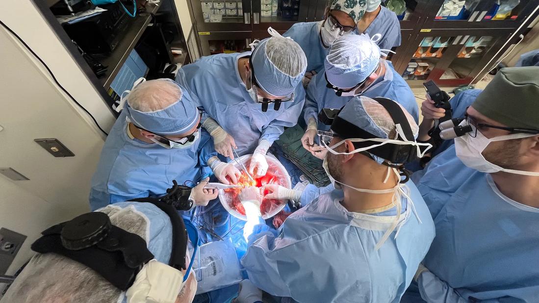
Image content: This image is available to view online.
View image online (https://assets.clevelandclinic.org/transform/c2cda105-f974-4198-a739-403773676004/intestine-auto-transplant)
Surgeons operating on intestines
A 38-year-old patient presented to Cleveland Clinic with a retroperitoneal schwannoma encasing the superior mesenteric artery. Her tumor had been diagnosed five years prior at roughly 9.4 × 8.9 cm, but a previous resection attempt was aborted due to vascular involvement.
Advertisement
Cleveland Clinic is a non-profit academic medical center. Advertising on our site helps support our mission. We do not endorse non-Cleveland Clinic products or services. Policy
“In conventional surgery, tumors involving the root of mesentery or major abdominal blood vessels are considered unresectable because removing them would cut off blood flow and cause organ necrosis,” explains Masato Fujiki, MD, a liver-intestine transplant surgeon at Cleveland Clinic. “Theoretically, those organs could be replaced with intestinal allotransplantation, but that would require lifelong immunosuppression that would increase risk of infection, kidney damage and malignant disease.”
Over the five years between her diagnosis and presenting at Cleveland Clinic, the tumor continued to grow. By 2026, imaging measured it at 13.1 × 11.3 cm, and the mass had begun preventing her from eating.
“During the past couple of years, I had been discussing an approach — ex vivo tumor resection with intestinal autotransplantation — with some colleagues,” says Dr. Fujiki. “This is an operation that has only been performed about 30 times over the past couple of decades.”
Unlike conventional transplantation, intestinal autotransplantation uses the patient’s own intestine. The bowel is temporarily removed, the tumor is resected outside the body and the intestine is then reimplanted, eliminating the need for a donor organ or lifelong immunosuppression.
The procedure is believed to be the first reported intestinal autotransplantation performed in Ohio and is among only a small number of such operations reported in the United States.
“After multidisciplinary discussion with our surgical oncology colleagues, we determined that this patient was well suited to the autotransplant approach,” says Dr. Fujiki. “This operation required expertise that crosses traditional surgical specialties. Our surgical oncology teams have tremendous experience with the Whipple procedure, while our transplant team brings expertise in organ preservation, vascular reconstruction and intestinal transplantation. Bringing those capabilities together made this approach possible.”
Advertisement
The procedure included ex vivo tumor resection with a Whipple procedure, pancreaticogastrostomy, intestinal autotransplantation, superior mesenteric artery reconstruction using an interposition iliac artery graft and a splenorenal shunt.
“We divided the pancreas and bowel and cut the involved vessels, allowing us to remove the tumor and affected tissue as a single specimen,” explains Dr. Fujiki. “Once it was removed, we autotransplanted the patient’s small bowel and right colon.”
The surgical team began with a Whipple procedure and creation of a splenorenal shunt. The head of the pancreas, the entire small intestine and the right side of the colon were resected en bloc. The right internal iliac artery was harvested for a vascular graft by Ravi Ambani, MD. Matthew Walsh, MD, and Robert Naples, DO, performed the pancreaticogastrostomy, connecting the remaining pancreatic tail to the stomach. The iliac artery graft was connected to the remaining portion of the SMA to reconstruct arterial blood flow. Intestinal transplant surgeons Mohammed Osman, MD, and Mohamed Maklad, MD were integral members of the operative team and remained closely involved in the patient’s complex postoperative care and recovery.
On the back table, the team removed the pancreaticoduodenal complex while preserving the main trunks of the superior mesenteric vein and SMA, as well as the first jejunal artery. Removing the organs allowed the surgeons to see the tumor and involved vessels more clearly and perform a precise resection without the bleeding that would complicate surgery inside the abdomen.
Advertisement
The intestinal and right colon autograft was then implanted, and the SMA was reconnected. The superior mesenteric vein was connected end-to-end to the main portal vein to restore venous drainage.
The total operating time was just over 14 hours. Despite the procedure’s length, estimated blood loss was 300 mL, and the patient did not require a blood transfusion.
Final pathology confirmed a 15.5 cm retroperitoneal schwannoma. The surgical margins were negative, indicating complete tumor removal, and two lymph nodes were negative for metastatic disease.
Although the initial operation went well, a pancreatic leak led to serious complications and required a completion pancreatectomy about two months later.
“The patient spent three months in the hospital,” says Dr. Fujiki. “She has recovered well, is eating well and is becoming more active.”
“The ex vivo procedure offers a way to achieve complete tumor removal when conventional surgery is not possible,” says Dr. Fujiki. “It expands the options for patients whose tumors involve the root of mesentery or major abdominal blood vessels.”
However, awareness of ex vivo tumor resection with intestinal autotransplantation remains limited, as does the number of centers with the necessary experience. Only about 100 intestinal transplants are performed each year nationally. Cleveland Clinic performs approximately 10 to 15, making it one of the country’s higher-volume programs.
Dr. Fujiki plans to build a specialized program at Cleveland Clinic around ex vivo tumor resection with intestinal autotransplantation, giving clinicians a referral option for patients whose tumors might otherwise be considered inoperable. The goal, he says, is to ensure that patients with tumors involving major abdominal vessels are evaluated at an experienced center before all surgical options are ruled out.
Advertisement
Advertisement

Transplant centers vary substantially in how they assess, document and follow patients after allogeneic transplantation
Pre-operative SMA balloon occlusion helped reduce intraoperative blood loss in a high-risk procedure
Insights on bringing Cleveland Clinic even closer to becoming the best transplant enterprise in the world
Consensus statement aims to increase use of the perfusion technology and raise transplant volumes
Minimally invasive pancreas-kidney replacement reduces patient’s pain, expedites recovery
AHA recommendations for pretransplant evaluation, peritransplant and long-term management
First-ever procedure restores patient’s health

The agent-based model aims to improve prediction accuracy
 Search