Locations:

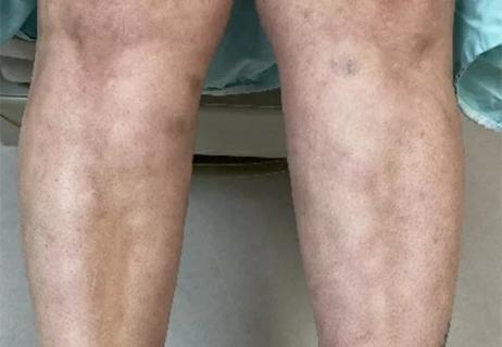
Patient’s symptoms included periorbital edema, fevers, weight loss
Video content: This video is available to watch online.
View video online (https://www.youtube.com/embed/UQeYCQfD_U8?feature=oembed)
Making the Puzzle Pieces Fit – VEXAS
A rare and sometimes fatal condition that was discovered as recently at 2020 can cause a constellation of symptoms so common that it can be difficult to home in on an accurate diagnosis.
In a recent video case study, Cleveland Clinic rheumatologist Adam Brown, MD, describes the case of a 71-year-old man who first sought help for symptoms that included fevers, night sweats and rash along with a 30-pound weight loss over three months. Periorbital edema — first unilateral and then progressing to bilateral — also was present. During examination, he was found to have low white blood count, low ANC, low hemoglobin with elevated mean corpuscular volume, but normal platelets.
Advertisement
Cleveland Clinic is a non-profit academic medical center. Advertising on our site helps support our mission. We do not endorse non-Cleveland Clinic products or services. Policy
He had neutrophilic dermatosis of his upper back. A CT scan of his chest, abdomen and pelvis revealed splenomegaly. A bone marrow biopsy revealed hyper cellularity.
“From the rheumatology perspective, when we have periorbital edema, the differential’s fairly narrow: Is it infection? Is it malignancy?” says Dr. Brown. “If those are ruled out, then the concern is something that can add tissue from rheumatologic perspective, which is actually not very broad.”
The patient was given high-dose steroids and felt better. The fever and both resolved, and cytopenias improved. He still didn’t have a diagnosis, however, and during tapering of prednisone after he left the hospital, a new symptom appeared: ear swelling, pain and redness.
The patient finally sought care at Cleveland Clinic, where clinicians diagnosed him with VEXAS — short for “vacuoles, E1 enzyme, X-linked, autoimmune, somatic mutation syndrome.” The condition was identified in 2020. The UBA1 gene is implicated in the condition, which is tends to affect patients 70 or older and more men than women.
To learn more, listen to Dr. Brown’s video case presentation.
Advertisement
Advertisement
How to use? Consider starting during the acute attack and seek patient preferences for chronic use

Distinct MRI signature includes lesions beyond the corpus callosum, features predictive of vision and hearing loss

Despite less overall volume loss than in MS and NMOSD, volumetric changes correlate with functional decline

It’s time to get familiar with this emerging demyelinating disorder
Early experience with the agents confirms findings from clinical trials
Despite advancements, care for this rare autoimmune disease is too complex to go it alone
Treatment for scleroderma can sometimes cause esophageal symptoms
Could the virus have caused the condition or triggered previously undiagnosed disease?