Treating a patient after a complicated hernia repair led to surgical complications and chronic pain

A 55-year-old woman with a history of Crohn’s disease presented after having multiple hernia surgeries. The patient explained that in 2018, she saw a surgeon at an outside hospital who offered her a robotic-assisted abdominal wall reconstruction. During that surgery, the surgeon injured both lateral complexes. On postoperative day six, the patient re-herniated through one of the layers of the closure and had to go back emergently. The patient developed several abscesses requiring reoperation, and the surgeon repaired one of the injuries from before but missed one.
Advertisement
Cleveland Clinic is a non-profit academic medical center. Advertising on our site helps support our mission. We do not endorse non-Cleveland Clinic products or services. Policy
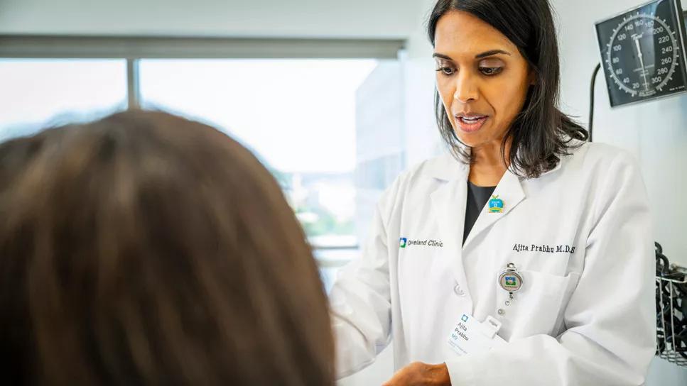
After two other medical centers saw the patient and refused to operate, the patient came to Cleveland Clinic in 2019 and saw Ajita Prabhu, MD, a general surgeon in Cleveland Clinic’s Digestive Disease Institute. Dr. Prabhu diagnosed the patient as having both her rectus muscles denervated and a left-sided hernia that had not been repaired at the time of take back.
The patient had nerve damage on both sides due to the lateral abdominal wall injuries. She also complained of chronic abdominal pain, worst in the left lower quadrant, and a bulging abdominal wall contour due to her defunctionalized abdominal wall. She also noted a chronic dull burn in her abdominal wall.
Dr. Prabhu felt the patient would benefit from a re-do abdominal wall reconstruction to repair her left-sided hernia, which was significantly symptomatic. The patient was informed that the surgery would not be able to restore her original abdominal wall contour or completely resolve the bulging and burning she is experiencing, since the damage from the original surgery was likely irreparable. However, Dr. Prabhu anticipated that the left lower quadrant pain should improve along with the patient’s overall abdominal wall function and quality of life (QoL).
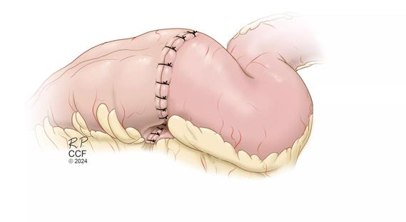
Dr. Prabhu performed the redo complex abdominal wall reconstruction surgery in February 2020 without postoperative complications. Robotic surgery was not an option in this case due to the multiple prior operations and anticipated hostile operative field, so the case was performed through an open approach.
Advertisement
In March 2020, the patient had a virtual visit with Dr. Prabhu for a follow-up. The patient noted that she was still having chronic pain, as expected per the preoperative discussion. However, her left lower quadrant pain and abdominal wall function were significantly improved. In May 2020, the patient came in for a follow-up and still complained of chronic pain. Due to the ongoing chronic pain, which was anticipated preoperatively, she was also referred to pain management.
In August 2022, the patient came back for a follow-up once more, with a new complaint of left lower quadrant pain and a minimal hernia recurrence where there appeared to be a small mesh fracture on CT imaging. Given that the hernia was not clinically apparent on exam and was < 2 cm in diameter on imaging, Dr. Prabhu discussed with the patient that an attempt at repair would be of significant scope for minimal improvement, and the patient agreed. She noted that her quality of life has significantly improved since the initial reoperation performed by Dr. Prabhu. Dr. Prabhu notes that at this point, they are monitoring the patient once a year with a computed tomography (CT) scan. At the patient’s more recent appointment in August 2023, she came back for an in-person follow-up appointment and noted that while the pain has improved somewhat, it is still present. Overall, she is pleased with her outcome and feels that the repair has significantly improved her quality of life and allowed her to maintain her employment.
Advertisement
The component separation technique that was used by the original surgeon is a complicated one, says Dr. Prabhu. “It’s a technique that wasn’t developed until around 2012, and it wasn’t applied to robotic surgery until years later. Even today, most attending surgeons were not trained to do this operation in residency. In the United States, few medical centers are doing this operation routinely enough to teach their residents how to do it. So, surgeons aren’t learning this in residency, and they end up learning about the technique a bunch of different ways — watching videos, going to conferences, telementoring, etc. The unintended consequence of this variability in training is that if surgeons attempt these operations without a strong understanding of the techniques, abdominal wall anatomy and familiarity with potential complications, they can cause inadvertent but devastating injuries for patients which may have lifelong repercussions. In our practice, we are seeing similar complications to the one presented here weekly.”
Cleveland Clinic does approximately 500-600 of these cases annually. Along with Dr. Prabhu, six other surgeons essentially perform the surgery on a full-time basis. A fellowship program at Cleveland Clinic teaches this type of operation, and the department includes seven surgeons who routinely perform the surgery at three locations.
“I would say that by the time our residents leave our program, they have a significant exposure to abdominal wall reconstruction techniques because it’s an integral part of their residency,” says Dr. Prabhu. “Our center performs the highest volume of these procedures in the country. We tend to skew more towards open surgeries simply because we tend to see more patients who are not candidates for minimally invasive approaches, but we still do enough robotic cases that we’re comfortable with them as well.”
Advertisement
Dr. Prabhu notes that a lot of what’s done in the hernia division is done utilizing a team approach. Many of the surgeons operate and also see patients in clinic on the same days, so they’ll often bounce ideas off of each other. The team also presents cases formally in a monthly hernia conference where we can review images and clinical scenarios together with our colleagues from other institutions to make sure we tailor the plans for the most complex cases.
“I don’t think that can be overstated enough because even though I treated and operated on this specific patient, everybody on our team has expertise so we always have the opportunity to review preop planning and operative approaches with each other, and we do this fairly frequently, especially for complicated cases,” says Dr. Prabhu. “We philosophically manage things the same way, and we’ve built our team so that there are redundancies. Every patient gets the same high level of care, no matter the surgeon.”
“For me, I think this case highlights the importance of being comfortable with and being able to operate in the gray areas,” explains Dr. Prabhu. “What I mean by that is many surgeons will say, ‘If I don’t have a binary outcome of, I’ve won, or I’ve lost, then there’s no point in operating.’ We see so many patients at Cleveland Clinic who are coming to us because they’ve been told no somewhere else. We have a ton of routine surgeries, but I think at the end of the day, the real reason that we’re here is to help those patients in the gray areas. Outcomes don’t need to be binary, and if we can help patients and improve their lives even a little bit, that’s a win for me.”
Advertisement
She continues, “In the case of this particular patient, we knew that her abdominal wall contour abnormality and chronic pain would persist, but we felt that we had a good shot at improving her quality of life. We have certainly accomplished that here despite the symptoms she continues to suffer from due to her initial complications.”
While Dr. Prabhu notes that there is a slight chance that the patient will need additional surgery in the future, she feels that the chance of that is not exceedingly high as she has continued to follow the patient with imaging and the hernia has remained stable over the past two years.
Advertisement

Retrospective analysis looks at data from more than 5000 patients across 40 years

Study utilizes prospective data from the RISK cohort

Nationwide research underscores the importance of individualized treatment

The Integrated Program aligns IBD care and research across Cleveland Clinic locations

Case study in diagnosing ACNES and finding the right surgical fix

Diet has a profound impact on how the intestine functions

Findings support the safety of the technique

Strong patient communication can help clinicians choose the best treatment option