Locations:

Scleromyxedema may cause extracutaneous symptoms
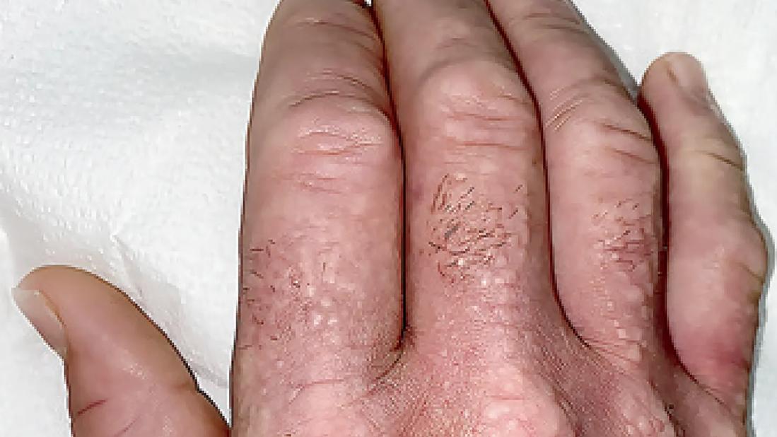
Image content: This image is available to view online.
View image online (https://assets.clevelandclinic.org/transform/ce9a3867-a9b2-4aa1-ac46-b4d3be76666b/Schleromyxedema-Hand)
waxy papules on his hands, with associated skin thickening and finger flexion contractures on hand
By Soumya Chatterjee, MD, and Anthony P. Fernandez, MD, PhD
Advertisement
Cleveland Clinic is a non-profit academic medical center. Advertising on our site helps support our mission. We do not endorse non-Cleveland Clinic products or services. Policy
A 53-year-old man presented to the rheumatology clinic with a three-year history of an itchy rash, Raynaud’s phenomenon, dysphagia and a burning sensation in his hands.
Physical examination was notable for firm, greasy papules across his forehead that led to the formation of glabellar grooves (A).

Image content: This image is available to view online.
View image online (https://assets.clevelandclinic.org/transform/111d397a-2d38-444d-8221-f1dac2dce09d/Scleromyxedema-Forhead)
A. Papules across the forehead led to the formation of glabellar grooves.
There were waxy papules on his hands with associated skin thickening and finger flexion contractures (B., above). Similar skin changes were seen on his nose, lips, ears, trunk and feet. There was no telangiectasia or calcinosis.
Sensory neuropathy was present in his hands, arms and face. Tests of thyroid function were normal. Serum protein electrophoresis with immunofixation identified an IgG-λ monoclonal gammopathy, and a bone marrow biopsy was normal.
A subsequent skin-biopsy sample obtained from the right side of the neck showed dermal spindle-cell proliferation, thickened collagen fibers, fibrosis and perivascular inflammation (C, hematoxylin and eosin stain), as well as increased dermal mucin deposition (D, colloidal iron stain). A diagnosis of scleromyxedema was made.

Image content: This image is available to view online.
View image online (https://assets.clevelandclinic.org/transform/47db125a-a0b0-41d8-ac03-0207422ad16f/Scleromyxedema-Stain-Slide)
C: A skin biopsy sample from the neck showed dermal spindle-cell proliferation, thickened collagen fibers, fibrosis and perivascular inflammation.

Image content: This image is available to view online.
View image online (https://assets.clevelandclinic.org/transform/bdd2fe67-d1ac-4d6b-83a4-f6febb34a2ec/Scleromyxedema-Blue-Slide)
D. Increased dermal mucin deposition.
Scleromyxedema is a primary cutaneous mucinosis typically associated with a paraproteinemia. This type of sclerosing skin disorder may cause extracutaneous symptoms, as was seen in this patient.
Although infusions of intravenous immune globulin provided minimal relief initially, treatment with lenalidomide resulted in abatement of symptoms and reduction in paraproteinemia after four months.
This article was originally published in The New England Journal of Medicine, Nov. 23, 2023.
Advertisement
Advertisement

Treatment insights from the ninth recorded EMPD case in 50 years
Treatment strategies require understanding of pathomechanisms

Multidisciplinary teams bring pathological and clinical expertise

First-in-human phase 1 trial induced loss of function in gene that codes for ANGPTL3

Major study demonstrates importance of having a multidisciplinary approach to treatment for large, locally advanced tumors

Researchers Assess Real-Life Experiences of Patients Treated Outside of Clinical Trials

Early, individualized diagnosis and comprehensive management key to preserving fertility

Key considerations when diagnosing and managing severe hyponatremia