Locations:

Education, prevention strategies and monitoring serves this at-risk group
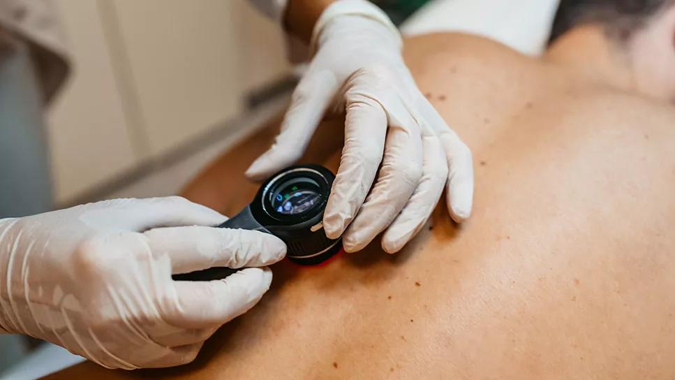
Image content: This image is available to view online.
View image online (https://assets.clevelandclinic.org/transform/d72f9312-862d-42ef-a533-ef6b1260f668/doc-Looking-at-Moles-1437090427)
Squamous cell skin cancer
Advertisement
Cleveland Clinic is a non-profit academic medical center. Advertising on our site helps support our mission. We do not endorse non-Cleveland Clinic products or services. Policy
You take care of a 72-year-old male with granulomatosis with polyangiitis (GPA). He presented with severe disease involving the lung and kidney nine years ago, requiring admission to the intensive care unit. Within the first year, he experienced one relapse after maintenance therapy was held for a serious infection. Since that time, he has remained in remission on azathioprine. He experienced chronic renal insufficiency from his initial presentation and now has a baseline creatinine 1.9 ml/dL with eGFR 35 ml/min, which has been stable. His quality of life has been excellent and in prior discussions he has expressed his wish to remain on azathioprine with the goal of reducing his risk of relapse.
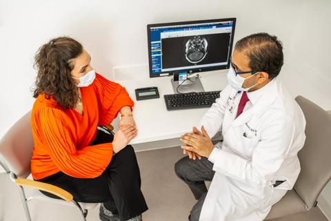
As a young man, the patient worked outdoors and had significant sun exposure, with later development of numerous actinic keratoses (AK). He now sees a dermatologist every three months to monitor for skin cancers, which he develops on a regular basis. His dermatologist contacts you to discuss his management.
Skin cancer is the most common cancer in the United States, with current statistics suggesting that one in five Americans will develop skin cancer in their lifetime.1 Squamous cell carcinoma (SCC), the most frequent form of skin cancer, typically arises from AK, which consist of aberrant epidermal keratinocytes that develop in response to prolonged ultraviolet (UV) radiation exposure. AK are considered the initial lesion in a disease continuum that may progress to SCC.
Early detection plays a key role in minimizing the spread of skin cancers and providing the patient with the widest range of options for treatment. People of all skin tones can develop skin cancer, with higher risk being seen in patients who have increased UV exposure from sunlight or use of indoor tanning beds, blond or red hair, and skin that burns easily.
Advertisement
Treatment with immunosuppressive medications also represents an important risk factor for skin cancer development, making skin cancer of even greater relevance to patients with rheumatic disease. The largest body of supportive evidence for this association comes from organ transplant recipients, for whom the risk of skin cancer is up to a hundred-fold higher than the general population.2-4
The mechanism through which immunosuppression increases skin cancer development is likely multifactorial, with reduction of host defense mechanisms that allow the body to detect and eliminate abnormal cells playing a prominent role. Immunosuppressive medications may also hasten the progression of AK to SCC and may themselves have properties that influence mutagenic potential. In transplant patients, the duration and dosage of immunosuppressive medications have been found to correlate with skin cancer risk as well as the type of immunosuppressive drug.
Skin cancer development has been well-described with conventional immunosuppressive agents such as cyclophosphamide, azathioprine, methotrexate, mycophenolate and calcineurin inhibitors. Although it is less clear that a similar association exists with biologic agents and small molecule inhibitors, taking a cautious approach that any medication that impacts host defense may increase skin cancer risk offers an opportunity to protect our patients against this common malignancy.
For patients who do develop non-melanoma skin cancers, withdrawal of immunosuppressive medications is often not an option in cases where the patient has a rheumatic disease where these are necessary to control the underlying inflammatory process. In most instances, it may also not be a good option to switch to a different agent in a patient who is doing well, as making a change will not eliminate the risk of skin cancer, the alternative also may not control the underlying disease to the same degree, and it will be associated with its own toxicities that could pose a greater threat than skin cancer where there is a careful program of monitoring and early detection.2-4
Advertisement
In most instances, it may also not be a good option to switch to a different agent in a patient who is doing well, as making a change will not eliminate the risk of skin cancer, the alternative also may not control the underlying disease to the same degree, and it will be associated with its own toxicities that
could pose a greater threat than skin cancer where there is a careful program of monitoring and early detection.
The first step toward reducing skin cancer risk for immunosuppressed patients comes in recognizing this association and providing proactive patient education. Preventive strategies include avoidance of indoor tanning beds. When outdoors, clothing should be used to cover as much exposed skin as possible. To protect skin not covered by clothing, it’s a good idea to wear a wide-brimmed hat and sunglasses with UV protection, and apply a broad-spectrum, water-resistant sunscreen with an SPF of 30 or higher.
In addition to protection against skin cancer, these strategies have additional benefit in rheumatic diseases associated with photosensitivity and where patients are receiving medications that can increase the risk of sunburn (NSAIDs, sulfonamides, methotrexate are examples). Regular use of sunscreen is important but often underutilized by immunosuppressed patients.
An ever-increasing number of skin care products and cosmetics incorporate a sunscreen, making this easier to include in daily life. While older patients may have already sustained solar skin damage many years ago, reducing further injury is beneficial. For younger patients, early adoption of sunscreen use will provide later benefits.
Advertisement
Opportune times to provide reminders about solar protection include office visits going into summer months and when the patient mentions a planned vacation to a sunny location; it should be emphasized, though, that UV exposure occurs outside regardless of the weather conditions. If a patient comes to a clinic visit with a sunburn, it provides an occasion to discuss with them that this represents skin damage and increases the potential for skin cancer.
Monitoring for skin cancer is also essential. Self-checks looking for changes in size, shape or color of a skin lesion, new lesions or a sore that doesn’t heal can be valuable in early detection. Emphasizing that immunosuppressed patients should receive an annual skin assessment by a dermatologist is also important. For those who have demonstrated a propensity to develop skin cancers, more frequent dermatology visits may be necessary to detect early cancers and monitor evolving AK lesions.
This patient exemplifies the risk of skin cancer that can be seen in immunosuppressed individuals. He sustained significant cutaneous solar damage in his youth with the development of AK later in life. Following the addition of immunosuppression that was lifesaving in the management of his GPA, he is now developing skin cancers, which was discussed with his dermatologist. In shared decision making with the patient, we concluded that it was in his best interests to remain on azathioprine, which has been otherwise well tolerated, and which has controlled his disease activity.
Advertisement
He continues to maintain skin-protective measures and receive regular dermatology assessments. Skin cancer is a common and possibly underappreciated risk in immunosuppressed people with rheumatic disease. Patient education is important, as the use of preventive and monitoring strategies can lessen the risk of skin cancer development and allow detection of lesions at an early
point where these may be most effectively managed.
Advertisement
First-ever U.S. population-level retrospective analysis reveals many patients with systemic mastocytosis need faster intervention

Evidence suggests empathy influences immune function and well-being

Going beyond simple referrals to streamline patient access

Cleveland Clinic group works toward earlier diagnosis of this rare disease

Pembrolizumab does not improve outcomes, but immunotherapy may still offer benefit

Unraveling the TNFA receptor 2/dendritic cell axis

Nasal bridge inflammation, ear swelling and neck stiffness narrow the differential diagnosis

Genetic testing at Cleveland Clinic provided patient with an updated diagnosis