Locations:

A reliable and reproducible alternative to conventional reimplantation and coronary unroofing
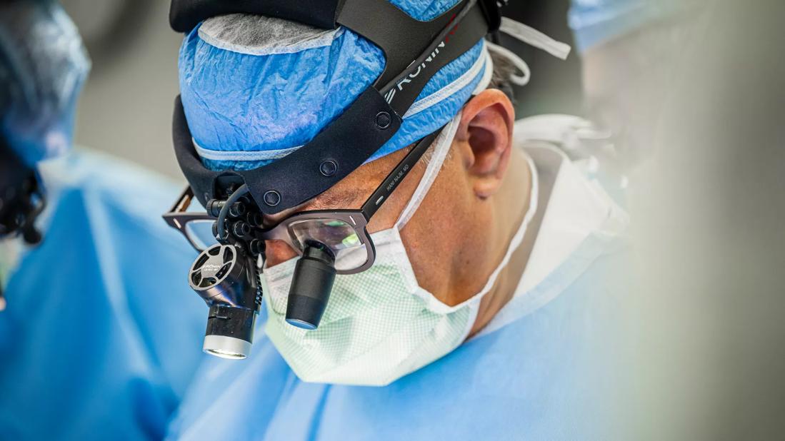
Image content: This image is available to view online.
View image online (https://assets.clevelandclinic.org/transform/9734ea10-c276-4feb-b995-0bd66f851d78/CCC_4891058_05-29-24_034_SCG)
Dr. Najm in the operating room wearing surgical loupes
Cobrahead reimplantation, a novel technique introduced by Cleveland Clinic cardiothoracic surgeon Hani Najm, MD, offers an alternative to conventional repair methods for anomalous right coronary artery (RCA) arising from the left coronary sinus with an intramural segment. The strategy has resulted in improved postoperative symptoms and a wider, more sustainable anastomosis.
Advertisement
Cleveland Clinic is a non-profit academic medical center. Advertising on our site helps support our mission. We do not endorse non-Cleveland Clinic products or services. Policy
A team of cardiothoracic surgeons describes the method and its outcomes in a case series published in The Multimedia Manual of Cardiothoracic Surgery.
RCA is a subvariant of anomalous aortic origin of coronary arteries (AAOCA) with an estimated prevalence ranging from 0.025% to 0.25% and representing less than 3% of congenital coronary anomalies. AAOCA is associated with sudden cardiac death in children and young people, and surgical intervention is indicated for patients with symptoms or evidence of ischemia.
Patients with this coronary defect typically have a short intramural course, conventionally repaired with a coronary unroofing technique. However, this method presents several challenges, according to Dr. Najm, who is Chair of Pediatric and Adult Congenital Heart Surgery.
“The intramural segment of the coronary artery sometimes runs behind the valvular pillar, extending from one sinus to the other, making unroofing difficult. Other variables that can lead to a complicated unroofing include a thick segment or a course lower than the aortic valve inter-commissural pillar. Detaching the valve becomes necessary to complete a full unroofing,” Dr. Najm says.
Reimplantation is an alternative option that enables surgeons to avoid unroofing altogether. “Instead of allowing the artery to dive into the aortic wall, we cut it off and reimplant it where it’s supposed to go,” he says. Still, reimplantation poses technical issues, specifically related to the right angle orientation in the traditional technique. This can lead to suboptimal takeoff from the ascending aorta, especially in a small coronary artery.
Advertisement
Dr. Najm adopted the cobrahead technique, which creates a larger anastomosis at the connection site with the aortic wall, to address this issue. “Typically, the diameter of the coronary artery is not more than 2 to 3 millimeters, and there is a possibility of getting a stenosis at the ostium of the implantation,” he notes. “However, if we do the cobrahead technique, then it will be at least double the size of that artery, which yields itself to a better longer-term anastomosis.”
The idea to utilize the cobrahead technique, a mainstay in coronary bypass surgery proximal anastomosis, came to him during a challenging unroofing procedure. “It occurred to me—why don’t we just reimplant it instead? I have done it this way in thousands of bypass surgeries,” he says, adding, “In surgery, there is a long tradition of innovating existing techniques when you find an opportunity for improvement.”
Dr. Najm and the team have completed eight cases using this technique. The patient characteristics and postoperative outcomes for seven of these patients are documented in the case series and detailed as follows.
Patients’ ages range from 16 to 63, and symptoms included chest pain, syncope and cardiac arrest. Median bypass and cross-clamp times were 75 and 37 minutes, respectively. Each patient had normal left ventricular and right ventricular function, with no reported aortic valve regurgitation, as determined by postoperative echocardiogram evaluation. Comparing pre- and postoperative catheterizations (5/7, 71%) revealed an improvement in the instantaneous wave-free ratio (IFR) of 0.18. At the time of follow-up, all patients showed an improvement in their symptoms.
Advertisement
“All of the patients have done well and with no complications. Our data show improved results even six months after the surgery, and that’s how we can confidently advise patients to return to their daily lives without restrictions to their activities.”
Center protocol, developed by Dr. Najm, guides eligibility for the surgery. A patient must present with two of the three factors: symptoms, a positive ischemia test, as indicated by a PET-CT scan, or a positive IFR.
In rare situations when a patient is asymptomatic, tests are negative, and the anomaly is detected incidentally, Dr. Najm notes, “We would operate if we believed there is a life-threatening risk.”
Dr. Najm emphasizes the importance of detailed imaging, including a preoperative CT scan and an angiogram, to provide visualization of the artery branches, inform surgical planning, and guide expectations for patients.
The operative steps are outlined below and also captured in a video created by the team.
Advertisement

Image content: This image is available to view online.
View image online (https://assets.clevelandclinic.org/transform/33460086-a3c9-43c2-ae14-2e9c651406eb/Intramural-RCA_technique-figure-2_23-HVI-3852632)
Key aspects of the cobrahead reimplantation technique. (A) A punch hole in the right coronary sinus with suture ligation of the distal end of the artery as it dives into the wall. (B) Spatulation of the distal end of the right coronary to create a cobra head. (C) Wide direct anastomosis of the coronary artery to the aorta.
Precision implantation and preservation of the artery are critical, as kinking and twisting of the artery can threaten the blood supply.
“This technique needs to be done by an experienced surgeon, but otherwise, we have shown that technique is reliable, it’s reproducible and gives us good results.”
Advertisement
Advertisement

A new CME opportunity in Chicago, May 15-16

Innovative hardware and AI algorithms aim to detect cardiovascular decline sooner

Experts advise thorough assessment of right ventricle and reinforcement of tricuspid valve

Reproducible technique uses native recipient tissue, avoiding risks of complex baffles

Program will support family-centered congenital heart disease care and staff educational opportunities

Case provides proof of concept, prevents need for future heart transplant

Pre and post-surgical CEEG in infants undergoing congenital heart surgery offers the potential for minimizing long-term neurodevelopmental injury

Science advisory examines challenges, ethical considerations and future directions