Case provides proof of concept, prevents need for future heart transplant

A 22-year-old woman was born with a double outlet right ventricle with cross-cross orientation of the atrioventricular valves and superior-inferior ventricles (also known as upstairs-downstairs ventricles). A large perimembranous inlet ventricular septal defect was also present along with a straddling mitral valve and bilateral superior vena cava.
Advertisement
Cleveland Clinic is a non-profit academic medical center. Advertising on our site helps support our mission. We do not endorse non-Cleveland Clinic products or services. Policy
In childhood, she had undergone pulmonary artery banding, atrial septectomy, a bidirectional Glenn and lateral tunnel fenestrated Fontan, all of which were performed at Cleveland Clinic. In recent years, she had developed venovenous collaterals, which required multiple coil embolizations and progressive subaortic right ventricular outflow tract obstruction (90 mm Hg).
The patient had been well enough to participate in martial arts and boxing during high school. But recently, she found that exercise triggered cyanosis of her lips, dyspnea and sometimes syncope, prompting her current presentation to Cleveland Clinic.
When she presented for surgical consultation, she underwent multiple diagnostic tests, including cardiac catheterization, cardiac MRI and various tests for other organs. These tests confirmed a high gradient obstructing the right ventricle. Her left ventricle was dilated with normal function, and she also had liver and kidney dysfunction.
"We discussed options for repair, including novel division of her heart that would return it to two ventricles, a procedure called a "biventricular conversion," says Hani Najm, MD, Chair of Pediatric and Congenital Heart Surgery. This option would include an aortic valve implantation.
“She decided to have a mechanical valve and agreed to proceed with an innovative conversion. The time was right because she was well enough to withstand a major surgery and keep her own heart. Had we waited, she might ultimately have needed a heart and a lung transplant," he explains.
Advertisement
A crucial first step in this novel procedure was Dr. Najm’s advance fashioning of a custom-made extra-anatomical graft. A size 23-mm mechanical On-X® aortic valve was attached to the stented end of a 28-mm Relay®Pro non-bare metal stent. The custom prosthetic was further lengthened with a 28-mm Dacron Hemashield graft (Figure 1).

Image content: This image is available to view online.
View image online (https://assets.clevelandclinic.org/transform/da9152f9-bd1d-4dba-9b8b-e4f9c8ce0188/Biventricular-conversion_Conduit)
Figure 1. Extra-anatomical valved conduit was key to complex ventricle conversion
"My goal was to create a conduit that turned around from the apex of the left ventricle, making a U-turn to meet the ascending aorta. Similar conduits were successfully used from the left ventricle apex to the descending aorta but not the ascending aorta. Creating a U-shaped conduit gave me confidence in the approach," explains Dr. Najm.
The conduit was sewn into the apex of the left ventricle using bovine pericardium sutured 1 cm from the flaring edge of the stent.
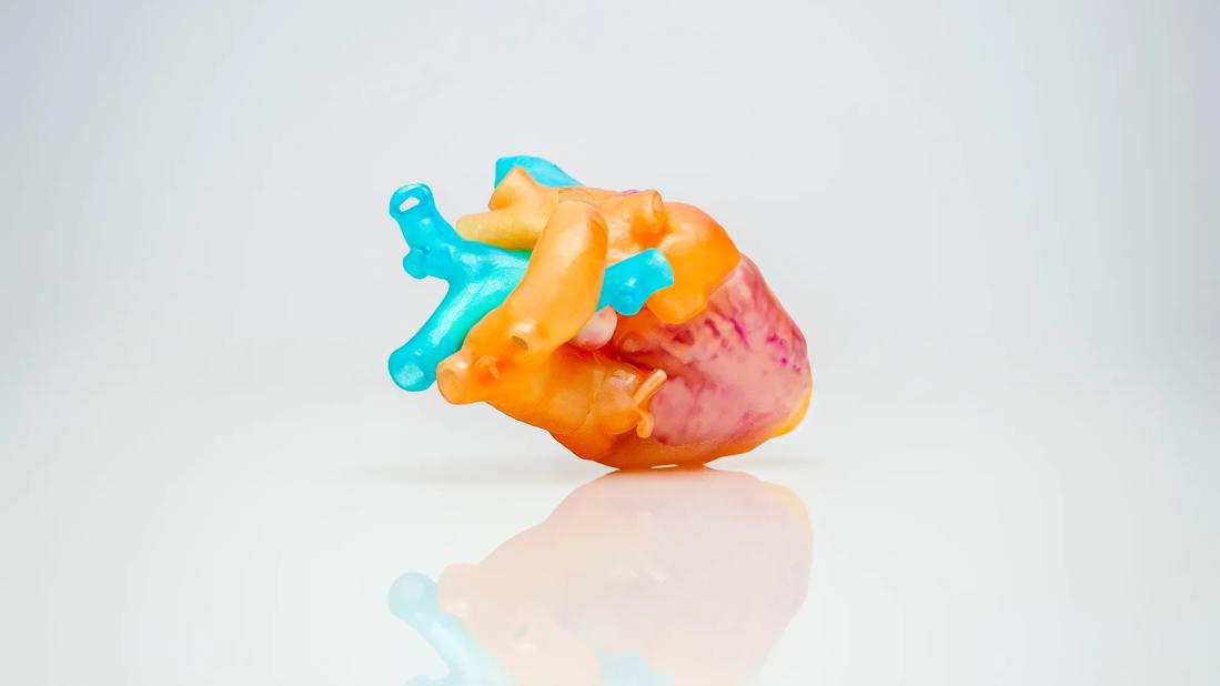
"Surgical innovation requires preparation,” he adds. For this case, such preparations included advanced imaging to create a 3D-printed model of the patient's heart (Figure 2). Dr. Najm and the team also conducted a presurgical virtual-reality walkthrough to visualize the reconstruction better and prepare for the challenges they might face in the operating room.

Image content: This image is available to view online.
View image online (https://assets.clevelandclinic.org/transform/76676b9c-ec54-40b9-9977-220c489ebd0a/najm-patient-3d)
Figure 2. 3D-printed model of patient's heart
This complex reconstructive surgery took approximately eight hours and required a highly skilled team. The key surgical steps included the following:
Advertisement
The coronary translocation was performed using the Cabrol technique with an 8-mm Dacron tube. Deficits in the aortic root were patched, the aortotomy was closed and the aorta was transected more distally.
"As the aorta was converted to a neo-pulmonary artery after the arterial switch, it was then attached to the left pulmonary artery," says Dr. Najm. "To prevent a pulsatile cavopulmonary anastomosis, a restrictive band was placed between the left pulmonary artery and the right-sided bidirectional Glenn.” (Figure 3).

Image content: This image is available to view online.
View image online (https://assets.clevelandclinic.org/transform/ac5320f9-231a-4990-be5a-3eb9b461c9fa/Biventricular-conversion_Upstairs-downstairs-Labeled)
Figure 3. A detailed rendering of the postoperative anatomy.
During the patient's six-week postoperative recovery, she developed diastolic dysfunction, which was successfully managed with diuretics and afterload reduction. This was not unexpected, according to Dr. Najm. Both ventricles had been pumping as a single ventricle for years. Therefore, this change will take time to adjust, he says.
Now, a year postsurgery, the patient is clinically asymptomatic and has returned to the gym. However, she can no longer practice martial arts because of the need for anticoagulant therapy. Echocardiography shows that her heart has adapted well to its new circulation. She will continue to be followed on an outpatient basis, but Dr. Najm anticipates no need for further surgery.
"This is proof of concept that the aortic valve and coronaries need not be on the usual outlet of the left ventricle," says Dr. Najm. "The one-and-a-half biventricular conversion alleviated our patient's subaortic right ventricular outflow tract obstruction, relieved her symptoms and improved her overall quality of life."
Advertisement
The surgery also precludes the patient from future heart and lung transplants. Dr. Najm advises, "Biventricular conversion should be considered, whenever possible, for individuals with early Fontan-related complications who are clinically well and have no overt organ failure."
Advertisement
Advertisement

Innovative hardware and AI algorithms aim to detect cardiovascular decline sooner

Experts advise thorough assessment of right ventricle and reinforcement of tricuspid valve

Reproducible technique uses native recipient tissue, avoiding risks of complex baffles

A reliable and reproducible alternative to conventional reimplantation and coronary unroofing

Program will support family-centered congenital heart disease care and staff educational opportunities

Pre and post-surgical CEEG in infants undergoing congenital heart surgery offers the potential for minimizing long-term neurodevelopmental injury

Science advisory examines challenges, ethical considerations and future directions

Updated guidance and a call to action