Locations:

Add AI to the list of tools expected to advance care for pain patients
Podcast content: This podcast is available to listen to online.
Listen to podcast online (https://www.buzzsprout.com/2243576/18152345-tech-driven-treatments-for-neck-back-pain)
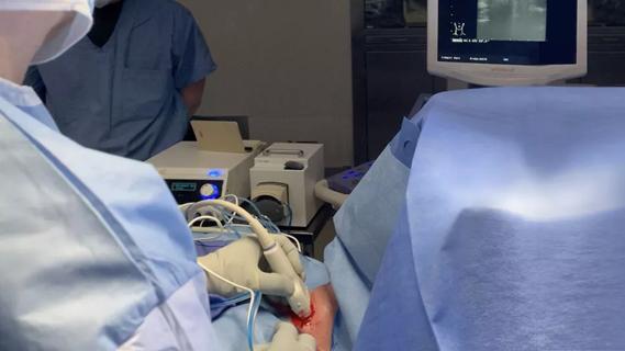
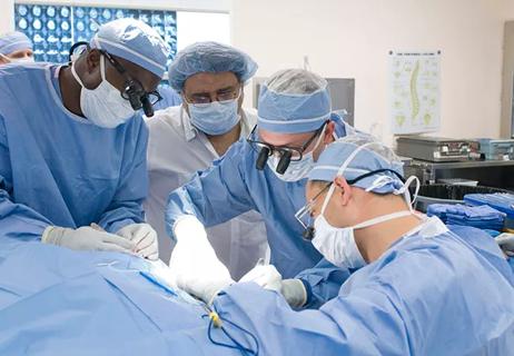
Technological advancements over recent decades have expanded treatment options for people with long-term or chronic pain conditions, and artificial intelligence (AI) promises to significantly expand on those innovations, says Trishul Kapoor, MD, a specialist in interventional pain management in Cleveland Clinic’s Neurological Institute.
Advertisement
Cleveland Clinic is a non-profit academic medical center. Advertising on our site helps support our mission. We do not endorse non-Cleveland Clinic products or services. Policy
“The biggest thing on the horizon is integration of artificial intelligence into current mechanisms of clinical practice,” says Dr. Kapoor. “With neuromodulation, we're … taking the journey that pacemakers took for cardiology, and we're just at the initial steps.”
In Cleveland Clinic’s Neuro Pathways podcast, Dr. Kapoor spoke to host Glen Stevens, DO, PhD, about technologies that are enabling pain management clinicians to improve the diagnostic process for patients and provide strategies to improve function and quality of life.
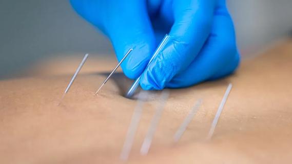
Chronic pain is notoriously complex, but AI, paired with neuromodulation technology, is expected to play an important role in enabling physicians to provide personalized and data-driven care.
“Technology is going to be a huge component of any clinical practice, and adoption and introduction to patients is going to be crucial,” says Dr. Kapoor. “It's just figuring out within the patient journey at what point which technology has the greatest impact, and knowing that there are various roles at different times.”
Dr. Kapoor discusses a variety of tech-related topics in the conversation, including:
Click the podcast player above to listen to the 30-minute episode now, or read on for a short, edited excerpt. Check out more Neuro Pathways episodes at clevelandclinic.org/neuropodcast or wherever you get your podcasts.
Advertisement
This activity has been approved for AMA PRA Category 1 Credit™ and ANCC contact hours. After listening to the podcast, you can claim your credit here.
Host Glen Stevens, DO, PhD: So I'm sure if you image an old guy like me, you're going to see lots of little things here and there, and I'm going to have some nonspecific complaints. I imagine it gets very complicated to figure out what's causing what. Talk to me a little bit about the technology that's now at your hand that you can use to help define if what you're seeing on the imaging study is correlating with my problem.
Trishul Kapoor, MD: In terms of the current technology that's available is... Our go-to gold standard is an MRI. Magnetic resonance imaging comes either in a closed format, which is a standard mechanism, or an open MRI if there are other restrictions based on patient claustrophobia or just body mass index. The basic mechanism for us is to look at an MRI of the lumbar spine, if that's the region of pain that's causing the issue, or looking at the cervical spine. Rarely, it ends up being a thoracic spine issue. Based on that, then we go on to look at other modalities, such as EMGs. So an electromyogram will correlate the findings within an MRI, or vice versa, to say where exactly the pathology could be, because the nerve itself can have a route all the way from your spine to an end terminal point, including the foot, the hand, whatever it may be. And anywhere along the way in its journey, it could potentially have a pathology.
Advertisement
Dr. Stevens: Any other diagnostic tools that you're using?
Dr. Kapoor: Outside of the normal sequencing, we're looking at new sequencing techniques that are not currently available, but research has shown things like 3D isotropic sequencing, which is a high-resolution image with isotropic voxels, as they call it, and it's basically representing a value in a three-dimensional regular grid. It allows us to create multi-planar reconstruction and helps improve visualization of the anatomy, especially in the spine. That's one thing.
The other thing is we realized that MRIs tend to be long and arduous, and so the question is, well, what can we do at a minimum to get away with constructing the other images? So we started looking into things like synthetic MRIs. This is a technique that allows us to generate multiple contrast views, T1, T2, STR imaging, from a single acquisition, which could potentially halve the scan time and provide quantitative T1, T2, and proton density maps for further analysis.
Advertisement
Advertisement

What the postoperative data show so far
Radiofrequency ablation significantly reduces symptom severity, shrinks nodules

Telehealth aids in treatment of fibromyalgia and median arcuate ligament syndrome
Program enhances cooperation between traditional and non-pharmacologic care
A surgeon’s perspective: three patient groups

CMR-CLIP outperforms general AI tools; may one day expand patient access to CMR

Collaboration with AI startup promises to reshape neurocritical care monitoring at scale

Collaboration includes clinical validation of predictive modeling tool, development of second-generation tool