Locations:





New studies clarify how sensory loss and nystagmus shape real-world visual function
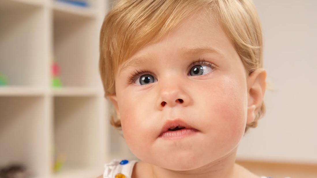
Image content: This image is available to view online.
View image online (https://assets.clevelandclinic.org/transform/6bb8d91c-caf4-4962-affb-1e014aff9558/strabismus-peds-2)
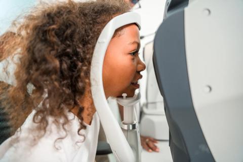
Child with strabismus
Most ophthalmologists think of amblyopia in terms of reduced visual acuity in one eye and impaired stereopsis. But those deficits alone do not explain how patients function in the real world.
Advertisement
Cleveland Clinic is a non-profit academic medical center. Advertising on our site helps support our mission. We do not endorse non-Cleveland Clinic products or services. Policy
“We are increasingly recognizing that amblyopia is not a monocular problem,” says pediatric ophthalmologist Fatema Ghasia, MD, Director of the Ocular Motility and Visual Neurosciences Lab at Cleveland Clinic Cole Eye Institute. “It’s actually a binocular disorder with a constellation of visual sensory deficits and eye movement abnormalities.”
Two recent studies from the Cole Eye Institute offer new insight into amblyopia. The findings show that fixation and saccadic abnormalities persist during binocular viewing and that nystagmus plays a major role in ocular motor behavior.
Video content: This video is available to watch online.
View video online (https://cdnapisec.kaltura.com/p/2207941/sp/220794100/playManifest/entryId/1_idxm4jtj/flavorId/1_5f3sgelj/format/url/protocol/https/a.mp4)
Video of a patient with strabismic amblyopia and nystagmus during binocular viewing. Note the coexistence of jerk nystagmus and strabismus. These abnormal eye oscillations can impair visual stability, limiting effective visual information acquisition and compromising both binocular visual acuity and the accuracy of saccades, which are often disconjugate and unequal between the two eyes.
Amblyopia affects an estimated 3% to 5% of the U.S. population, and delayed detection can result in lifelong visual impairment. Standard clinical testing remains largely monocular, even though most real-world visual function is binocular.
According to Dr. Ghasia, ocular motor abnormalities may help explain why some patients continue to struggle with reading, visual scanning and other visuomotor tasks despite standard treatment for amblyopia.
In a recent study published in the Investigative Ophthalmology & Visual Science special issue on nystagmus co-edited by Dr. Ghasia, Dr. Ghasia and colleagues used video-oculography to characterize fixation eye movement abnormalities in healthy controls (n = 64) and patients with amblyopia and/or strabismus (n = 97), both with and without nystagmus. The study provided new insights into the relationship between fixation stability and visual function.
Three findings were especially notable:
Advertisement
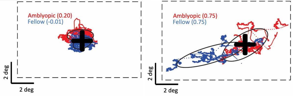
Image content: This image is available to view online.
View image online (https://assets.clevelandclinic.org/transform/3944650d-9169-4fd7-9644-a2c17114064c/abnormalities-amblyopia-inset)
Graphs show extent of fixation instability in both eyes when viewing with fellow eye (left) and amblyopic eye (right). The focal target is marked with a black cross. Fixational eye movement of the fellow eye is represented in blue; fixational eye movement of the amblyopic eye is represented in red.
These findings suggest that interocular differences in fixation stability could serve as objective biomarkers of amblyopia severity.
“From a screening perspective, we potentially can look at the interocular difference in fixation stability and identify a child more likely to have amblyopia,” Dr. Ghasia says. “That could be very valuable for young patients not yet able to read an eye chart. We could possibly detect amblyopia earlier so we can begin treating it earlier.”
In a second study, also published in Investigative Ophthalmology & Visual Science, Dr. Ghasia and colleagues used video-oculography to measure visually guided saccades (rapid eye movements made to peripheral targets) during binocular and monocular viewing in patients with healthy eyes (n = 21) and in patients with amblyopia and/or strabismus (n = 49).
The data revealed that some abnormalities tracked with amblyopia severity, while others were driven primarily by nystagmus.
Notably, the basic mechanics of saccade generation remained intact across groups. This suggests the deficits arise from abnormal sensory processing and disrupted binocular development rather than a motor abnormality.
Advertisement
“Studies often exclude patients with nystagmus or do not analyze them separately,” Dr. Ghasia says. “Now we know that nystagmus should not be ignored in studies of amblyopia and strabismus.”
Together, the two studies help clarify how amblyopia, strabismus and nystagmus each contribute to abnormal eye movements.
The key takeaways are:
These findings have important clinical implications. They suggest that ocular motor abnormalities in amblyopia are not a single entity.
These findings do not yet change routine clinical care, but they strengthen the rationale for incorporating eye movement analysis into future approaches to amblyopia diagnosis and follow-up.
“Eye tracking offers an objective, nonverbal, pediatric-friendly way to capture fixation and saccadic behavior,” Dr. Ghasia says. “This is important because current instrument-based screening tools primarily identify amblyopia risk factors, such as anisometropia and strabismus, but have limited specificity for detecting amblyopia itself. As a result, many children are referred for further evaluation despite not having amblyopia.”
Eye movement markers could improve specificity when used alongside conventional screening tools, she adds. Some eye-tracking platforms use animated targets to engage children as young as age 2, allowing clinicians to assess gaze behavior, fixation and saccades.
Advertisement
As eye-tracking technology becomes more portable and less expensive, its use outside research settings may become more feasible. Over time, this may support broader adoption of eye tracking for amblyopia detection and monitoring, particularly when paired with artificial intelligence.
“I am more and more convinced that eye movements can become useful clinical biomarkers for diagnosis and monitoring of amblyopia,” Dr. Ghasia says. “Now we know that nystagmus, which is an objective finding, can indicate worse outcomes for a patient with amblyopia or strabismus. Those are the people who likely will have more real-world visual impairment with both-eye viewing, even after treatment.”
Advertisement
Advertisement
Learn the latest recommendations in diagnosis, treatment and surveillance
The drug does not provide lasting disease control for most patients
A review of pathophysiology, risk factors, clinical presentation and treatment options
Registry data highlight visual gains in patients with legal blindness
Study is first to show reduction in autoimmune disease with the common diabetes and obesity drugs
It’s the first step toward reliable screening with your smartphone
Fixational eye movement is similar in left and right eyes of people with normal vision

Oral medication may have potential to preserve vision and shrink tumors prior to surgery or radiation
 Search