Locations:





Learn the latest recommendations in diagnosis, treatment and surveillance
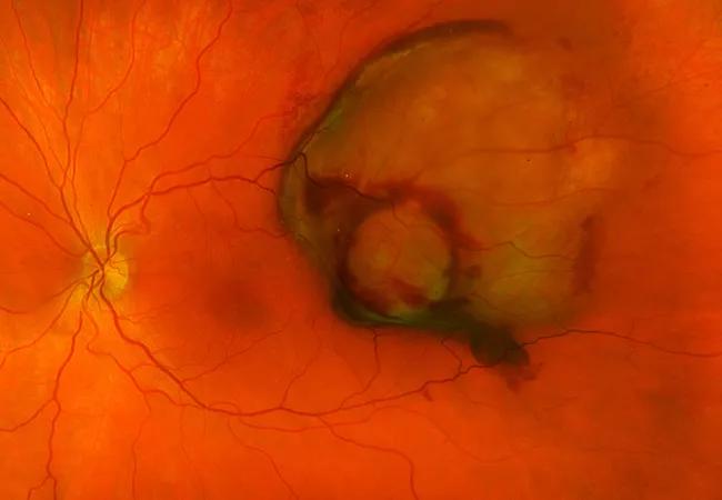
Image content: This image is available to view online.
View image online (https://assets.clevelandclinic.org/transform/0a8eb9ce-321b-41f6-b5a7-da50565daaba/21-EYE-2340059-CQD-Cured-fractions-of-uveal-melanoma_Arun-Singh-jpg)
uveal melanoma
Uveal melanoma, the most common type of ocular cancer in the U.S., affects approximately five in 1 million adults annually. The rare malignancy has various manifestations, but a structured approach to diagnosis, treatment and surveillance is possible and necessary, says Arun Singh, MD, Director of Ophthalmic Oncology at Cleveland Clinic Cole Eye Institute.
Advertisement
Cleveland Clinic is a non-profit academic medical center. Advertising on our site helps support our mission. We do not endorse non-Cleveland Clinic products or services. Policy
Updated National Comprehensive Cancer Network (NCCN) guidelines, released in 2026, provide a practical framework for managing suspicious lesions in the choroid and ciliary body and for tailoring follow-up based on metastatic risk.
“The value of the NCCN guidelines is that they are continuously updated to reflect emerging evidence and practical clinical experience,” says Dr. Singh, who presented the uveal melanoma recommendations at the 2026 NCCN Annual Conference.
While the complete document provides detailed flowcharts that guide clinical decision-making as well as full references supporting the recommendations, Dr. Singh says the points highlighted below are most relevant to ophthalmologists.
When a patient presents with a suspicious choroidal or ciliary body lesion, with or without visual symptoms, the recommended workup includes:
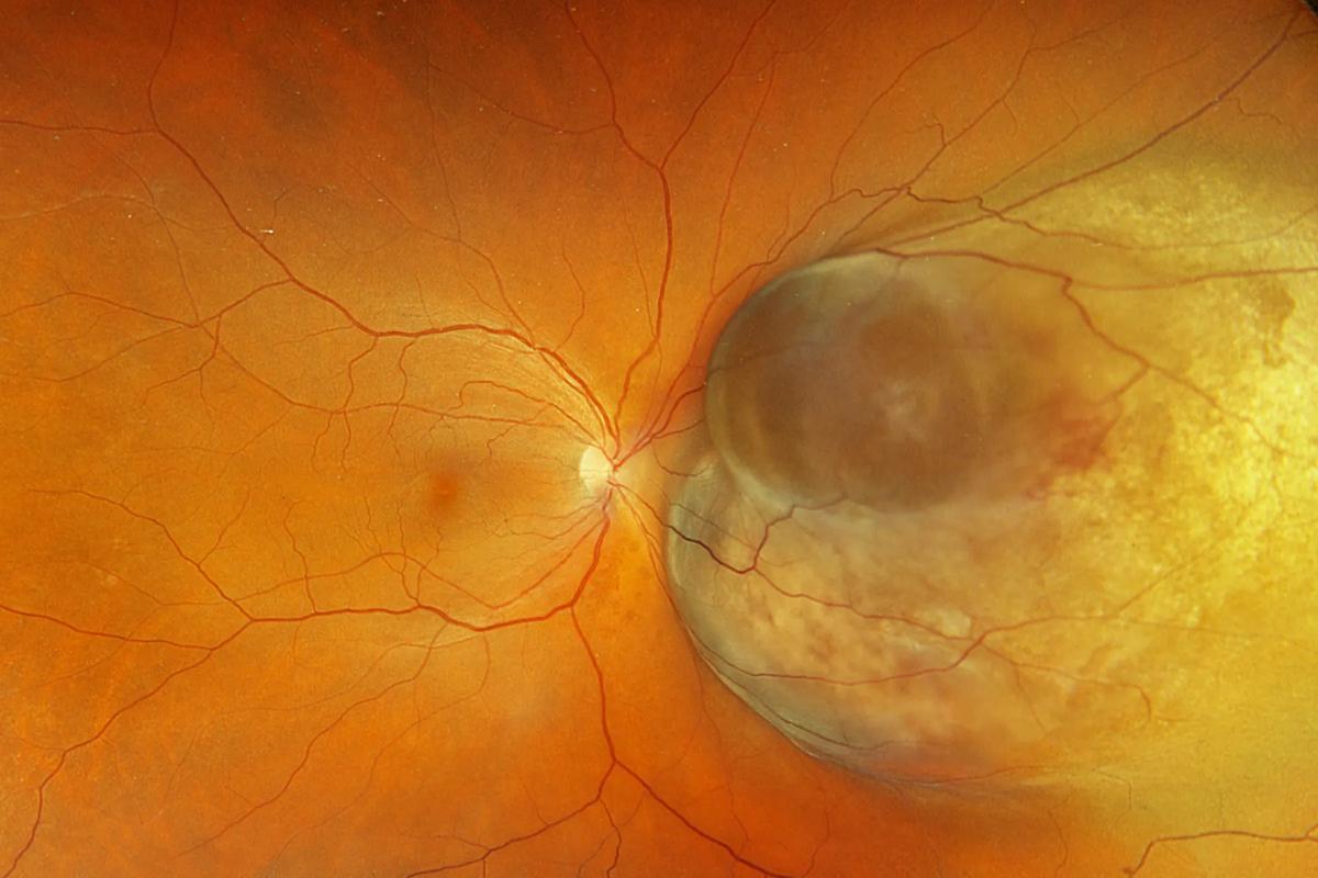
Image content: This image is available to view online.
View image online (https://assets.clevelandclinic.org/transform/d839ea26-b7a8-437f-869d-486e84d56c61/uveal-melanoma-1A)
Fundus appearance of a typical melanoma
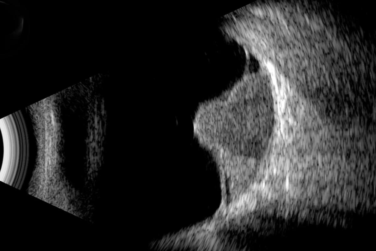
Image content: This image is available to view online.
View image online (https://assets.clevelandclinic.org/transform/f8186a6d-2fee-4243-895a-3ac248202efe/uveal-melanoma-1B)
Characteristic mushroom shape of uveal melanoma on ultrasound B scan
For small indeterminate lesions, close observation is appropriate. Reassessment is typically recommended every two to six months initially, then annually if stable. Features that increase concern for melanoma include:
Advertisement
Once a lesion is clinically diagnosed as uveal melanoma, additional evaluation should include:
Primary treatment selection depends largely on tumor dimensions, although location, extraocular extension, visual potential and patient preference also are factors.
For tumors 5-19 mm in diameter and less than 2.5 mm thick, treatment options include:
For tumors 19 mm or less in diameter and 2.5-10 mm thick, treatment options include:
For tumors more than 19 mm in diameter, more than 10 mm thick, or more than 8 mm thick involving the optic nerve, treatment options may include:
“These are not the only treatment options,” Dr. Singh notes. “Management must be individualized. In selected patients, observation or enrollment in a clinical trial may be the most appropriate course.”
The new guidelines also introduce a novel approach to metastatic risk stratification and surveillance.
Advertisement
“All uveal melanomas are not the same,” Dr. Singh says. “We must monitor them differently depending on their size and molecular profile.”
Tumors with low risk of distant metastasis include T1 lesions and those with favorable molecular features, such as:
Tumors with intermediate risk of distant metastasis include T2 and T3 lesions and those with one or more of these molecular features:
Tumors with high risk of distant metastasis include T4 lesions and those with one or more of these molecular features:
Recommended surveillance (e.g., contrast-enhanced MRI, CT abdomen and pelvis, abdominal ultrasound, CT chest, chest X-ray) varies by risk level:
“Metastases mostly occur in the liver, but they also can involve the lungs, bone, skin and other soft tissues,” Dr. Singh says. “As preventive and metastasis-directed therapies evolve, identifying which patients need closer surveillance will become even more important.”
Following the NCCN guidelines offers several benefits for both patients and clinicians, notes Dr. Singh.
Advertisement
“As ophthalmologists, we want to provide care that is grounded in the best available evidence,” he says. “The guidelines help us do that by offering expert-driven recommendations that standardize care, facilitate access to treatment and give patients resources that empower them to participate in their care.”
Advertisement
Advertisement

New studies clarify how sensory loss and nystagmus shape real-world visual function
The drug does not provide lasting disease control for most patients

A review of pathophysiology, risk factors, clinical presentation and treatment options

Registry data highlight visual gains in patients with legal blindness
Study is first to show reduction in autoimmune disease with the common diabetes and obesity drugs

It’s the first step toward reliable screening with your smartphone

Fixational eye movement is similar in left and right eyes of people with normal vision

Oral medication may have potential to preserve vision and shrink tumors prior to surgery or radiation
 Search