Locations:


Collaboration with AI startup promises to reshape neurocritical care monitoring at scale

Study looked at mobility measures and safety

Types and presentation may differ from adults, but early recognition and intervention are just as key

Study tests potential for a more accurate treatment predictor
Advertisement
Cleveland Clinic is a non-profit academic medical center. Advertising on our site helps support our mission. We do not endorse non-Cleveland Clinic products or services. Policy

Study uses molecular and clinical stratification to help guide patient selection

Phase 3 STELLAR trial underscores role of molecular stratification in glioma care

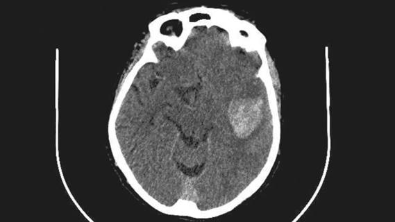
Early identification of temporal encephaloceles can improve surgical decision-making

New research highlights serious risks and the critical need for earlier advance care planning
Academia, industry and government leaders develop consensus priorities

Current diagnostic and treatment strategies can help improve focus, productivity and quality of life
Advertisement
Advertisement