Locations:

An overview of case reports and management
Advertisement
Cleveland Clinic is a non-profit academic medical center. Advertising on our site helps support our mission. We do not endorse non-Cleveland Clinic products or services. Policy
Nonthrombotic pulmonary embolism (NTPE) is uncommon and can be difficult to diagnose. My colleagues and I recently reviewed this complete or partial occlusion of the pulmonary vasculature resulting from inorganic particulate matter and foreign bodies in Chest. The below images demonstrate accidental embolization from catheter tips and guidewires.
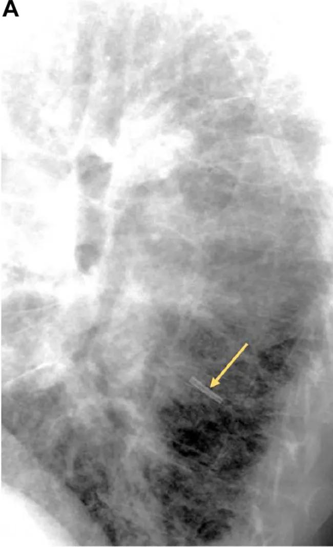
Image content: This image is available to view online.
View image online (https://assets.clevelandclinic.org/transform/b422e4a7-0996-4f4e-bd67-e70ba2c4e8d2/18-PUL-4584-Mehta-NPTE-Inset-Image-01-650pxl-width_jpg)
This lateral chest radiograph shows an embolized central venous catheter (CVC) tip in the left lower lobe sub- pulmonary artery branch. Intensivists commonly use CVCs for venous access. Potential complications include phlebitis, central line-associated bloodstream infections and catheter fracture with or without downstream embolization. Tip fracture and embolization like the example above can occur with any type of catheter, sometimes due to “pinch-off” syndrome, when a catheter placed in the subclavian vein gets wedged between the clavicle and the rib, leading to compression, transection and embolization.
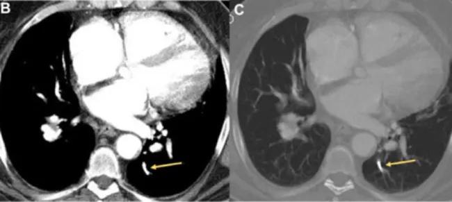
Image content: This image is available to view online.
View image online (https://assets.clevelandclinic.org/transform/b7c9017b-59d5-4250-9e5c-2403af10b7d9/18-PUL-4584-Mehta-NPTE-Inset-Image-02-650pxl-width_jpg)
These contrast-enhanced axial CT images (B, soft tissue window; C, bone window) at the level of the ventricles show an embolized catheter tip in the left lower lobe posterior sub-segmental artery.
NTPE has also been reported to occur when removing the guidewire or during catheter exchange. Catheter fragments have been known to lodge in the pulmonary artery, hepatic or brachiocephalic veins, superior vena cava, right atrium and right ventricle.
The below image demonstrates the gold standard of management for this type of NTPE: percutaneous retrieval via the femoral route. This isn’t always possible, and thoracotomy is an option. The image below shows (A) a contrast-enhanced chest CT with a linear radiopaque guidewire in the distal right main pulmonary artery extending into the proximal right lower lobe artery and right interlobar. The angiogram on the right (B) shows cannulation of the right pulmonary artery and fluoroscopic guided retrieval of the guidewire using a snare.
Advertisement
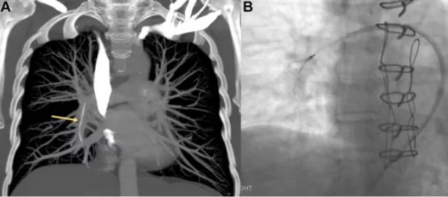
Image content: This image is available to view online.
View image online (https://assets.clevelandclinic.org/transform/73f1b698-ecda-43c5-bfa9-446381bc85f5/18-PUL-4584-Mehta-NPTE-Inset-Image-03-650pxl-width_jpg)
This is a rare occurrence, and patients may present with a wide spectrum of signs and symptoms from none to severe. Accurate diagnosis requires extensive knowledge of imaging modalities and postprocessing tools. Future posts will review other macroscopic emboli like radiation seeds and cement, microscopic emboli like ethiodized oil and silicone embolus and talc granulomatosis.
Cleveland Clinic coauthors include Sanjay Mukhopadhyay, MD, staff, Department of Anatomic Pathology, Derick Asah, DO, resident, and Subha Ghosh, MD, staff, Department of Diagnostic Radiology.
Dr. Mehta is staff in the Department of Pulmonary Medicine.
Images and text republished with permissions from Elsevier. Originally published in Chest.
Advertisement
Advertisement

An ICU pharmacist explains the impact of delirium on ICU patients and why there is a need for more research and improved screening tools

The Center for Environment, Place and Health Research connects clinicians with mapping tools and other real-world data resources to help guide medical insights

Exploring the true prevalence of AATD among Americans and why so many remain undetected

Rapid growth and collaboration have advanced the Lung Nodule Program, but patient engagement barriers persist

A physician's perspective on confirmation imaging, diagnostic confidence and choosing the right robotic platform for patients

A look into recent developments in the diagnosis of LAM and its clinical presentation

Takeaways from the most recent annual meeting centered around clinical advances, AI integration and professional development

Recent breakthroughs have brought attention to a previously overlooked condition