Locations:





Quantitative B-scan findings may offer a noninvasive way to assess vascular inflammation
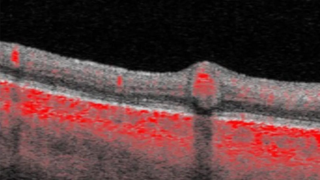
Image content: This image is available to view online.
View image online (https://assets.clevelandclinic.org/transform/0b7ca542-ceb1-4db6-b766-3d1a9a43e26c/OCT-scan-retina-vasculitis)
Layers of the retina highlighted in red
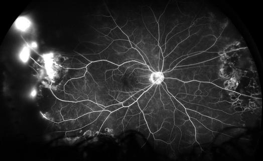
Fluorescein angiography (FA) remains the gold standard for clinically evaluating retinal vasculitis. FA can reveal vascular leakage, making it valuable for detecting active inflammation and guiding treatment. However, FA is invasive, requiring intravenous injection of dye, and many patients with uveitis need repeated studies over time to monitor their disease.
Advertisement
Cleveland Clinic is a non-profit academic medical center. Advertising on our site helps support our mission. We do not endorse non-Cleveland Clinic products or services. Policy
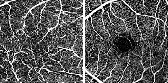
As retinal imaging has advanced, optical coherence tomography (OCT) and OCT angiography (OCTA) have transformed how clinicians assess retinal structure and perfusion. While OCTA offers a noninvasive method of evaluating blood flow, it cannot detect leakage from inflamed vessels like FA can. As a result, ophthalmologists still rely primarily on FA to assess vasculitis.
A team at Cleveland Clinic Cole Eye Institute is exploring a new way to assess vasculitis — by using quantitative OCT and OCTA to look at structural changes in inflamed vessels and surrounding tissue. The team’s recent study published in Ophthalmology Retina has revealed measurable differences that occur during active inflammation, such as increased retinal thickness near the inflamed blood vessel.
“These findings point to a new, noninvasive way to visualize and quantify vascular inflammation with OCT and OCTA instead of FA,” says lead author Yuka Mizuno, MD, a research fellow at the Cole Eye Institute. “This suggests that physicians may be able to objectively estimate a patient’s vasculitis activity without injecting dye.”
The study assessed 87 eyes from 50 Cole Eye Institute patients (mean age 43) with inflammatory retinal disease due to sarcoidosis, lupus, Crohn’s disease and other causes of uveitis. Researchers obtained OCT and OCTA B-scan data and compared it with FA imaging, searching for vascular and perivascular structural changes in areas of vessel inflammation.
Data from the study confirmed a correlation between vasculitis activity and structural changes in the retina.
Advertisement
Between active and inactive phases of retinal vasculitis, significant changes were noted in:
In addition, OCT B-scans showed unique hyporeflective and hyperreflective structural changes around inflamed vessels.
“Among our biggest revelations were those perivascular changes, which had not received much attention until now,” Dr. Mizuno says. “We were particularly surprised by how prominent the changes were around veins. Our other data quantitatively confirmed the physical expansion of vessels and surrounding tissues during active inflammation. The retina directly above the vessel bulges (increased elevation ratio) while the ONL below is compressed and thinned (decreased thinning ratio). It was also fascinating to find that the expansion of the vessel diameter was more pronounced vertically than horizontally, likely due to available space toward the vitreous cavity.”
Although structural changes can occur in tandem with leakage, the study found that those markers of inflammation do not always align. In some cases, FA indications of vascular leakage appeared unchanged while OCT findings showed clear perivascular swelling and other tissue changes around the vessels.
Advertisement
These observations suggest that vascular inflammation cannot always be fully characterized by leakage alone. Structural alterations in the inflamed vessels and surrounding tissues may become detectable on a different timeline than FA abnormalities.
“As clinicians, we tend to think of fluorescein leakage as the defining sign of active vasculitis, but inflammation is more complex than a single imaging feature,” says the study’s senior author Sumit Sharma, MD, a vitreoretinal surgeon and uveitis specialist at the Cole Eye Institute. “What OCT and OCTA B-scans allow us to see is the structural response of the vessel and surrounding tissue. In some eyes, those changes are evident even when the FA picture looks relatively stable. That is why it is so important to evaluate these patients from multiple angles.”
Used together, FA and OCT may provide a more complete picture of disease activity than one imaging modality alone, he says. This more comprehensive assessment may help physicians better understand subtle changes over time and refine treatments.
Structural changes apparent on OCT not only provide a new perspective on vascular inflammation, but also present a new method of noninvasive monitoring that could reduce the burden on patients, says Dr. Mizuno.
However, this monitoring technique is not ready for widespread clinical use.
“Currently, analysis requires manual reviewing or measurement of each B-scan, which is too complex for routine clinical practice,” she says. “For patients to truly benefit, we need to develop AI technology and software that can automatically detect specific changes and provide an intuitive assessment of activity. We are currently working on this.”
Advertisement
Advertisement
The advanced stage of diabetic retinopathy is among the most challenging for retinal surgeons

How to screen for and manage treatment-triggered uveitis
Researchers to study retinal regeneration in zebrafish with new grant from National Eye Institute

Leukocoria is the most common symptom of this rare childhood eye cancer
AI tool matches manual grading and may improve consistency in retinal research
Watch for sudden unilateral vision loss without pain
Prematurity is linked with poorer outcomes of retinal surgery in adulthood
A primer on sustained release options
 Search