Locations:





Digital subtraction myelography unveils often-overlooked but highly treatable underlying defect
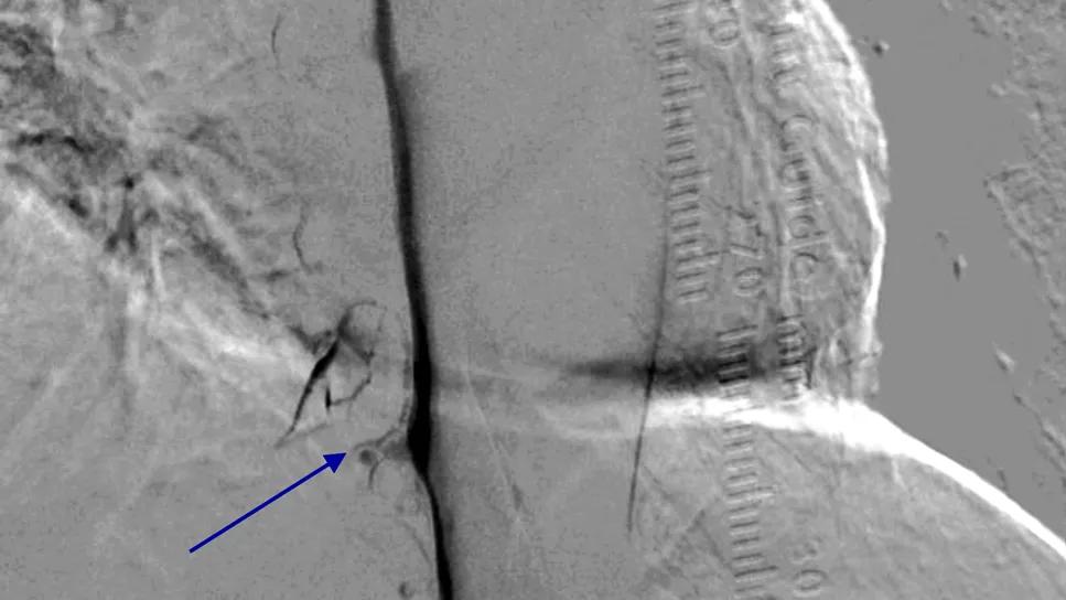
Image content: This image is available to view online.
View image online (https://assets.clevelandclinic.org/transform/8f3c3002-cee8-4bd1-be9e-c7bd4667ba45/remove-overlying-bone-to-isolate-contrast-flow-1)
scan of brain anatomy with a blue arrow marking a point of interest
In March 2025, a 63-year-old woman with a history of migraines went to a primary care clinic for severe headaches marked by pain radiating down the right side of her neck. She described the pain as increasing when she leaned forward, worsening with activity and subsiding with rest. She obtained no relief from over-the-counter pain relievers. A CT of the head was ordered but showed no abnormal findings. She was prescribed sumatriptan.
Advertisement
Cleveland Clinic is a non-profit academic medical center. Advertising on our site helps support our mission. We do not endorse non-Cleveland Clinic products or services. Policy
The patient returned in May 2025 reporting chronic headaches that had worsened in severity since their onset three months earlier and which were interfering with her ability to work. Coughing, sneezing and laughing caused the pain to intensify. Sumatriptan and a nonprescription acetaminophen/aspirin/caffeine combination provided only minimal relief. Allergy-aggravated symptoms were suspected, and daily prophylaxis with fluticasone and cetirizine was recommended.
The following month, the patient sought an opinion from a local neurologist. A brain MRI was requested. The MRI report noted a constellation of findings consistent with intracranial hypotension and a cerebrospinal fluid (CSF) leak. She was referred to a local neurosurgeon, who advised conservative management and to consider undergoing an epidural blood patch if her symptoms didn’t improve.
Dissatisfied with these recommendations, and after nearly a year of debilitating symptoms, the patient eventually arranged to see Cleveland Clinic endovascular surgical neurointerventionalist Mohammad Ammar Abdulrazzak, MD, who had started a new clinic at Cleveland Clinic Mercy Hospital only a few months earlier.
About 50% of patients with intracranial hypotension have an underlying CSF-venous fistula. As a woman in her 60s, this patient fit the demographic profile of individuals most likely to be affected. This, along with review of the patient’s earlier MRI, prompted Dr. Abdulrazzak to suspect that a CSF-venous fistula was the cause of her intractable headaches. “We expanded her workup to include dedicated spine testing aimed at identifying CSF leakage in the spine region,” Dr. Abdulrazzak explains.
Advertisement
Specifically, Cleveland Clinic neuroradiologist David Mihal, MD, performed a CT myelogram. No clear fistula was identified. When the initial CT myelogram showed no leak, Drs. Abdulrazzak and Mihal reviewed the images together and elected to proceed directly to digital subtraction myelography. This test identified a CSF-venous fistula associated with the right T10 nerve sheath. At this time, the patient was in a high level of distress. She was experiencing severe symptoms of brain fog and cognitive impairment in addition to her intractable, debilitating headache.
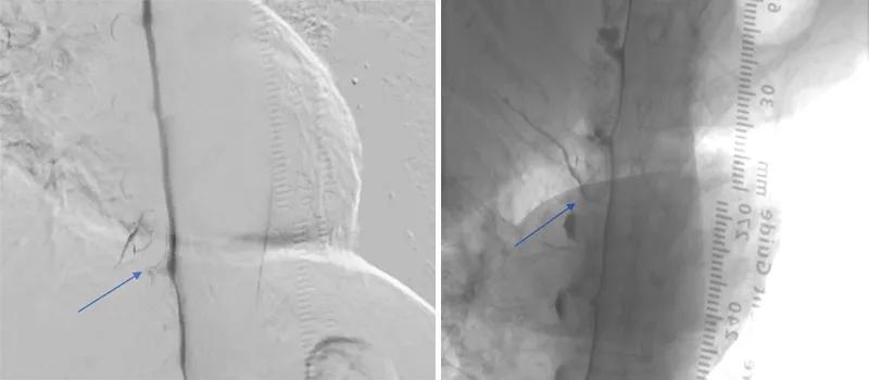
Image content: This image is available to view online.
View image online (https://assets.clevelandclinic.org/transform/3abf0790-f2bf-42cb-8d64-0ef3bb590643/subtracted-image-and-unsubtracted-inset1)
Digital subtraction myelography performed in a lateral decubitus position. The subtracted image (left) removes overlying bone to isolate contrast flow. The unsubstracted image (right) shows the same finding in anatomic context, with contrast escaping the thecal sac into a paraspinal vein at the right T10 nerve sheath. This is the fistula that was not visible on conventional imaging.
In April 2026, Dr. Abdulrazzak treated the patient with a minimally invasive endovascular procedure known as transvenous embolization. Through a percutaneous puncture at the site of the common femoral vein, a catheter was introduced into the venous system. A specialized balloon microcatheter was then navigated to the vertebral level where the CSF/vein connection was previously identified. Through the microcatheter, a liquid embolic agent was infused to fill and occlude the draining vein, disconnecting it from the CSF-filled diverticulum.
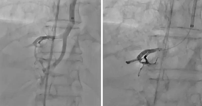
Image content: This image is available to view online.
View image online (https://assets.clevelandclinic.org/transform/a12452a4-2ecf-4ecd-94e8-0977b668ea22/transvenous-embolization-before-and-after-inset2)
Transvenous embolization. A balloon microcatheter was navigated into the segmental fistula draining vein, at right T10. Venography (left) confirmed its position. The balloon was then inflated to prevent reflux while the embolic agent was injected. The post-embolization image (right) shows the resulting embolic cast occluding the segmental vein, disconnecting it from the CSF-filled diverticulum.
When the patient awakened from anesthesia, her headache was gone. She was discharged home after an overnight hospital stay. At her first postoperative follow-up two weeks after the procedure, she reported that for the first time in over a year she could finally cough, sneeze and laugh without triggering pain.
A few weeks later she attended a concert by one of her favorite bands — an evening she had assumed was no longer possible — and invited family and friends who she said “had watched her lose the previous year to headache.” At her three-month follow-up in July 2026, her brain MRI was normal, with resolution of the imaging findings of intracranial hypotension. She now rates her headache 0/10 and her quality of life 10/10.
Advertisement
The incidence of spontaneous intracranial hypotension due to CSF-venous fistula has been reported as 2-3 per 100,000 person-years, although the precise prevalence of CSF-venous fistulas is uncertain.
There are multiple theories about how these fistulas form. The most likely explanation is that they are caused by a weakness in the spinal dura mater. For unknown reasons, the dura dilates, creating a CSF-filled space called a diverticulum that can connect with a nearby vein, resulting in low CSF pressure. Diverticula can occur anywhere along the spine.
“The hallmark of intracranial hypotension and a CSF-venous fistula is a debilitating orthostatic headache” Dr. Abdulrazzak notes. “It also can lead to cognitive impairment, anxiety or depression.” Potential headache triggers include leaning forward, sneezing, coughing and laughing.
In a supine position, affected patients do not have a headache. As soon as their head rises, the headache begins. Patients sometimes have difficulty expressing this and may say things like, “I can’t wait to go home and go to bed.” Dr. Abdulrazzak advises physicians to follow up on such comments with direct questions like, “Do you have a headache when you are lying flat? Does it start when you sit up?”
Due to the frequency of an underlying CSF-venous fistula, Dr. Abdulrazzak says patients with suspected intracranial hypotension should be referred to a tertiary center for care by a team of physicians familiar with the diagnosis of CSF-venous fistula, which can be challenging. In fact, the time from initial presentation to diagnosis is still commonly measured in months to years.
Advertisement
“The challenge,” he explains, “is recognition and high suspicion for the diagnosis. Patients can end up being treated for migraine headache or another headache syndrome. We now believe CSF-venous fistula is more common than previously thought. Awareness of this problem is very important to making the diagnosis and possibly pursuing treatment.”
Once the diagnosis has been made, transvenous embolization performed by an experienced interventional neuroradiologist can be done and typically has an immediate positive effect on the patient’s quality of life.
CSF-venous fistula returns in a minority of patients, but rarely at the same location. “When it returns, it can be treated in the same manner with the same excellent results,” Dr. Abdulrazzak concludes.
Advertisement
Advertisement

How effective management integrates multiple specialties and therapies

A close look at the growing array of options for episodic and chronic migraine

The selective D1/D5 dopamine agonist provides symptom relief and improves daily function.
5-year funding award supports cohort expansion and deep phenotyping of AD and related dementias

Highest response rates seen with stress-lowering techniques like yoga, meditation and mindfulness

Housing multidisciplinary clinicians in one leading-edge facility can transform GME along with patient care
How ongoing efforts will be bolstered by Cleveland Clinic’s new Neurological Institute building

Foundation model reveals latent risk structures in sleep physiology that elude conventional metrics
 Search