Locations:





Case: Presentation includes joint pain and neurological symptoms
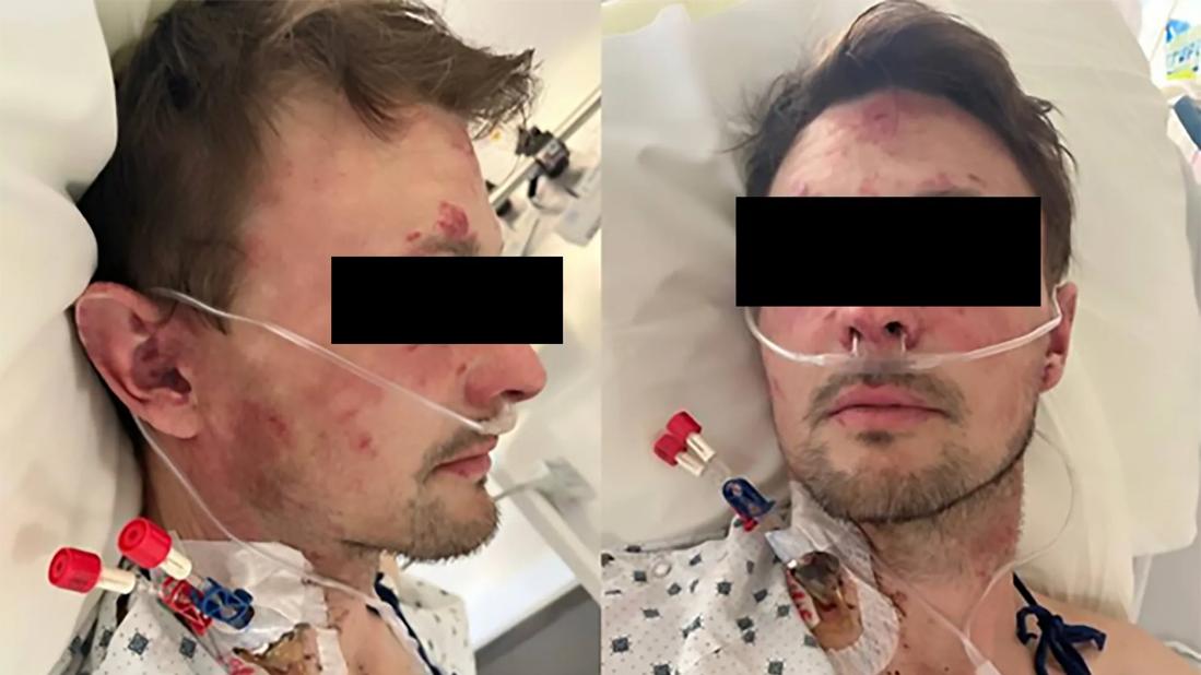
Image content: This image is available to view online.
View image online (https://assets.clevelandclinic.org/transform/4ec6564c-2b28-4fd5-9d5e-de65158c59ee/adam-brown-case-conference-lupus)
Two clinical images of a man with erythematous patches on his ace and ears.
By Yanint Raksadawan, MD, and Adam J. Brown, MD
Advertisement
Cleveland Clinic is a non-profit academic medical center. Advertising on our site helps support our mission. We do not endorse non-Cleveland Clinic products or services. Policy
A 35-year-old male with a history significant for chilblain lupus and childhood immune thrombocytopenic purpura (ITP) presented to the hospital with right perioral numbness, dysarthria and right arm numbness. On the day of admission the patient experienced a transient episode of perioral numbness accompanied by dysphagia, numbness in his right arm and leg and weakness in his right leg. He reported difficulty pressing the gas pedal with his right foot.
On review of systems, he also reported worsening pain in the joints of his hands. He otherwise denied new shortness of breath, chest pain, worsening leg swelling, dysuria and hematuria, as well as any history of blood clots.
In 2001, at age 11, the patient was diagnosed with ITP. He was treated with intravenous immunoglobulin and corticosteroids for two years without adequate response. He eventually received four doses of rituximab and has been in remission since.
In 2009, he began developing a malar rash and photosensitive rashes on his chest and arms. He started experiencing fatigue, arthralgia and Raynaud's phenomenon. A punch biopsy of a skin lesion was consistent with systemic lupus erythematosus (SLE). He was started on hydroxychloroquine, which was later discontinued because of adverse gastrointestinal effects.
In 2018, he established care with Cleveland Clinic Dermatology for persistent rashes on his hands, arms, face and extensor surfaces of the knees. A skin biopsy resulted in fi ndings consistent with chilblain lupus. He was treated with chloroquine, pentoxifylline and amlodipine.
Advertisement
Upon admission the patient’s vital signs were stable. Examination revealed extensive erythematous patches on the cheeks, eyelids, forehead and ears, sparing the nasolabial folds, as well as on the dorsal surfaces of all fingers, sparing the joints. Indurated, raised, skin-colored lesions were noted on both arms. The remainder of the physical examination was unremarkable, with normal joint and neurologic examinations.
Laboratory studies on admission were significant for a platelet count of 3,000 K/μL and hemoglobin of 8.5 g/dL. The white blood cell count and comprehensive metabolic panel were otherwise normal. The erythrocyte sedimentation rate and C-reactive protein were mildly elevated.
Given the initial presentation with neurologic symptoms, we had concerns for hemorrhagic stroke in the setting of severe thrombocytopenia. However, CT of the brain and CT angiography of the head and neck were unremarkable, aside from remote changes in the right frontal lobe attributed to a previous surgery following a motor vehicle accident.
Additional laboratory studies revealed low C3 and C4 levels, elevated LDH, an elevated reticulocyte count and low haptoglobin. A peripheral blood smear demonstrated true thrombocytopenia with numerous schistocytes (one to four cells per high-power field).
Given the thrombocytopenia with schistocytes, ADAMTS13 activity was checked and returned low. Rheumatologic serologies showed an ANA titer of 1:640 with positive anti-Smith, anti-RNP, antiribosomal RNP, anti-Scl-70, anti-dsDNA and anti-crithidia antibodies. Lupus anticoagulant, beta-2 glycoprotein antibodies and cardiolipin antibodies were all negative.
Advertisement
The patient’s history of ITP, biopsy-proven chilblain lupus, current skin changes with a distribution typical of SLE (malar rash sparing the nasolabial folds, lesions overlying the dorsum of the fingers sparing the joints), positive lupus serologies and low C3/C4, led to a diagnosis of active SLE.
Regarding his thrombocytopenia, the initial differential diagnosis included ITP because of his history and apparently active underlying SLE. However, with schistocytes identifi ed on the peripheral blood smear, concern for thrombotic microangiopathy (TMA) was higher. The differential diagnosis for TMA includes thrombotic thrombocytopenic purpura (TTP), antiphospholipid syndrome, and complement-mediated TMA/atypical hemolytic uremic syndrome.
His ADAMTS13 activity was low and his antiphospholipid antibodies were all negative, and he was diagnosed with TMA secondary to TTP.
With negative brain imaging studies, his initial neurologic symptoms were attributed to his TTP.
In summary, the patient was diagnosed with TTP based on his neurologic symptoms, microangiopathic hemolytic anemia, thrombocytopenia and low ADAMTS13 activity in the setting of active SLE, as evidenced by active cutaneous lesions and low C3/C4 levels.
He received five sessions of plasma exchange and was concurrently started on intravenous methylprednisolone 1 g daily for three days, followed by oral prednisone 60 mg daily with a subsequent taper.
Treatment with caplacizumab was initiated, along with rituximab 375 mg/m² weekly for four doses. Caplacizumab was discontinued after the patient developed epistaxis. An MRI of the brain performed six days after admission showed no hemorrhagic changes.
Advertisement
After initiation of treatment, the patient’s platelet count normalized within four days, and his neurologic symptoms did not recur. He was maintained on chloroquine, pentoxifylline, amlodipine and rituximab. His skin lesions were completely resolved after six months of treatment.
Cytopenias are a common manifestation of active SLE. It’s important to keep in mind life-threatening causes of anemia and thrombocytopenia in SLE, such as microangiopathic hemolytic anemia (MAHA). MAHA should be suspected in the setting of worsening anemia and thrombocytopenia.
Patients will have an abnormal haptoglobin and indirect hyperbilirubinemia with a blood smear demonstrating schistocytes. A few rheumatologic conditions have a higher risk of certain types of MAHA, SLE, anti-phospholipid syndrome, Sjogren's syndrome and scleroderma.1,2
In patients with SLE, various causes of MAHA should be investigated. Immune-mediated TTP is caused by immune inhibition of the enzyme ADAM metallopeptidase with thrombospondin type 1 motif 13, leading to the accumulation of large von Willebrand factor (vWF) multimers, triggering platelet aggregation leading to endothelial damage. Immunemediated TTP can be diagnosed in the setting of MAHA and low serum levels of ADAMTS13.
Another cause of MAHA in SLE is complement-mediated hemolytic uremic syndrome, caused by an acquired abnormality in the regulation of the alternative complement cascade on endothelial surfaces. Complement-mediated hemolytic uremic syndrome presents similarly to TTP and has the same laboratory fi ndings but normal ADAMST13 activity.
Advertisement
Rapid recognition and treatment are critical to treat either immune-mediated TTP or complement-mediated hemolytic uremic syndrome. Without treatment, these conditions lead to progressive neurologic decline, irreversible kidney damage and death. Patients are typically treated with plasmapheresis and high-dose glucocorticoids.
Considering the SLE, rituximab was used to treat the underlying condition, as Hematology felt this would be a reasonable treatment option for TTP, and we felt it would likely benefi t the SLE.
Caplacizumab is an approved therapy for TTP and works by inhibiting vWF from interacting with platelets to prevent the formation of microthrombi.
1. Babar F, Cohen SD. Thrombotic Microangiopathies with Rheumatologic Involvement. Rheum Dis Clin North Am. 2018
2. Kulkarni A, Vaughn K, Lacy C, Sultani T, Keshavamurthy C. Caplacizumab in Lupus-Associated Thrombotic Thrombocytopenic Purpura (TTP): Navigating Autoimmunity and Microangiopathy.
Cureus. 2025
Advertisement

Drug-free remission holds more than a year after treatment

CAR T-cell therapy may offer reason for optimism that those with SLE can experience improvement in quality of life.

Advancements lead to a new trial involving autoimmune disease

Lupus Clinic providers collaborate to advance treatment and understanding

A review of expanding options that hold new promise

Disease associated with higher risks for maternal and fetal morbidities
Early data show risk is 73% higher in patients with lupus, 40% higher in patients with rheumatoid arthritis
Cleveland Clinic’s Rheumatic Lung Disease program treats patients with complex conditions
 Search