Machine learning models assess intraoperative tissue perfusion

Image content: This image is available to view online.
View image online (https://assets.clevelandclinic.org/transform/310f57d1-05b9-428c-a68a-72b4ffa39d4d/Sarcoma-of-soft-tissue-feature)
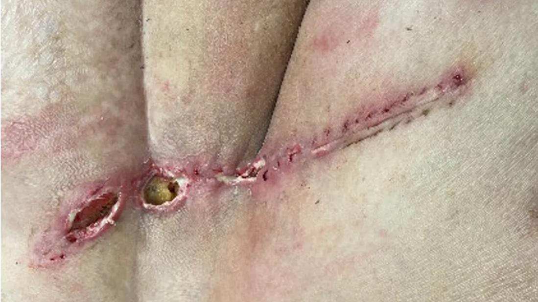
Incision after surgery to remove sarcoma
By Precious Oyem, MD, MSc; Lukas Nystrom, MD; and Nathan Mesko, MD
Advertisement
Cleveland Clinic is a non-profit academic medical center. Advertising on our site helps support our mission. We do not endorse non-Cleveland Clinic products or services. Policy
Soft tissue sarcomas are a rare group of heterogenous malignant tumors that are life-threatening but potentially treatable. If the tumor is localized, a combination of radiation therapy and surgical resection can be curative.
Wound healing complications are common after treatment of these tumors, especially when radiation therapy is used preoperatively. These complications occur in about 35% of all patients who undergo preoperative radiation (up to 43% if the tumor is in the lower extremity) and about 17% of those who undergo postoperative radiation.
The ability to predict these complications could help surgeons take steps to prevent them.
Initially, Cleveland Clinic’s Dr. Nystrom and several other orthopaedic oncologists at nationally reputable sarcoma institutions investigated transcutaneous oxygen measurements as a potential predictor of wound complications. Their study reported that transcutaneous oximetry was not predictive of wound complications. However, a noted limitation of the study was that the researchers sampled only a small portion of each wound.
When considering next steps in this investigation, a Cleveland Clinic team hypothesized that real-time assessment of tissue perfusion of the entire wound bed using a fluorescent dye, indocyanine green (ICG), would better predict wound healing complications after resection. ICG has been used for intraoperative assessment of perfusion in plastic surgery, for identification of lymph nodes and in assessment of cardiac function.
Advertisement
The sarcoma team at Cleveland Clinic launched a prospective study of patients having limb-sparing resection of soft tissue sarcoma, with or without radiation therapy, and with surgical incisions that were closed primarily. During surgery, dye was injected at various time points, and perfusion was captured using near-infrared fluoroscopy.

Image content: This image is available to view online.
View image online (https://assets.clevelandclinic.org/transform/969aede3-e040-4c31-82af-8e8c67091bac/Sarcoma-of-soft-tissue-inset1)
Intraoperative use of near-infrared fluoroscopy and intravenous ICG to reveal perfusion.
Data generated from the live image capture were collected. The patients were followed for six months and monitored for wound healing problems. Photographs of wounds were analyzed by mapping wound regions with their eventual outcome. Specific wound regions were correlated to the perfusion intensity/time curves attained intraoperatively.

Image content: This image is available to view online.
View image online (https://assets.clevelandclinic.org/transform/2bd1b3c6-b084-4872-9284-a0905b0a5375/Sarcoma-of-soft-tissue-inset2)
Top: Clinical photograph. Bottom: Graph of perfusion at varying regions of interest on the surgical wound (time, x axis; perfusion intensity, y axis).

Image content: This image is available to view online.
View image online ( https://assets.clevelandclinic.org/transform/b4f66243-6e7f-4756-a798-7985b2fbf107/Sarcoma-of-soft-tissue-inset3)
Top left: Intraoperative assessment of patient’s wound bed with respective perfusion/time curve (bottom). Top right: One-month clinical follow-up showing dehiscence in area of low intraoperative perfusion.
Machine learning models were used to analyze the preliminary data. Our models showed a high level of accuracy in predicting wound complications using data from the intraoperative perfusion/time curves alone (87%-94% accuracy).
This work, led by Drs. Nystrom and Mesko, has been presented at several national and international conferences. It won an award for best research at the 2024 J. Robert Gladden Orthopaedic Society’s research competition, where Dr. Oyem presented on behalf of Cleveland Clinic’s orthopaedic oncology team.
The powerful combination of tissue perfusion mapping and machine learning may open a gateway to a wealth of information. These predictive models can empower surgeons to proactively address potential wound complications intraoperatively. This could involve:
Advertisement
If the anticipation of wound healing complications are factored into the surgical planning process, it could enable surgeons to devise well-informed strategies to mitigate the risks. Furthermore, this capability could significantly aid patient counseling and expectation-setting.
Overall, the potential of this study is vast and paints a promising picture of the future of soft tissue sarcoma surgery, marked by a substantially reduced rate of wound complications.
Dr. Mesko is an orthopaedic surgeon and Section Head of Orthopaedic Oncology and Trauma at Cleveland Clinic. Dr. Nystrom is an orthopaedic surgeon who specializes in musculoskeletal tumors at Cleveland Clinic. Dr. Oyem is a resident in Cleveland Clinic’s Department of Orthopaedic Surgery.
Advertisement
Advertisement
Personalized reconstruction is an alternative to leg amputation or flail limb
Soft tissue pathologist discusses research into incorporating genomic data to improve risk stratification
Previous studies, trial aims and the potential role of immunotherapy
Innovative construct restores function after resection of softball-size chondrosarcoma
For kids with painful, growing lesions in the arm, leg or pelvis
Approach could help clinicians identify patients at an increased risk of progression who could benefit from more aggressive treatment
This rare disease usually manifests as a firm, painless mass that is growing
Cleveland Clinic researchers discover what drives – and what may halt – virus-induced cancer