Locations:

Personalized reconstruction is an alternative to leg amputation or flail limb
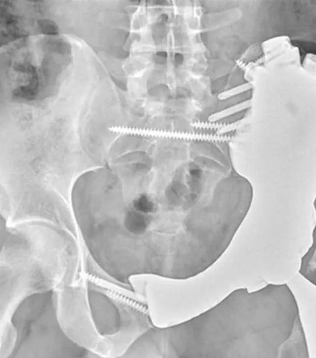
Image content: This image is available to view online.
View image online (https://assets.clevelandclinic.org/transform/68aa239c-4543-41b9-b9c4-807f86c9e9ab/3d-printed-pelvic-implant-feature)
X-ray of pelvis implant
A 29-year-old U.S. Army staff sergeant was referred to Cleveland Clinic after being diagnosed with chondrosarcoma in the left side of his pelvis.
Advertisement
Cleveland Clinic is a non-profit academic medical center. Advertising on our site helps support our mission. We do not endorse non-Cleveland Clinic products or services. Policy
According to Cleveland Clinic’s Section Head of Orthopaedic Oncology Nathan Mesko, MD, 10 years ago the only options after resecting the cancer would be to amputate the leg or leave a flail leg (connected to the pelvis only by soft tissue). In this case, Cleveland Clinic’s sarcoma team opted to replace the removed bone with a custom 3D-printed implant.
“Today we can get rid of the cancer, preserve the leg and restore the patient’s function,” Dr. Mesko says. “3D printing is common for creating surfaces on orthopaedic implants that mimic cancellous bone, fostering bone ingrowth. Now 3D printing entire custom-fit implants is becoming more common at high-volume sarcoma centers like ours.”
Using scans of the patient’s pelvis, the surgical team worked with engineers to create specialized guides to help precisely resect the cancer. Next they designed and printed a titanium implant to replicate the removed pelvis and fit tightly into the remaining geometry. The scans also helped Dr. Mesko map out every aspect of the surgery, including incision sites, soft tissue attachment sites and screw trajectory angles.
The entire process, including getting necessary approvals, took about eight weeks. Dr. Mesko then performed the 10-hour surgery to remove the sarcoma and affected pelvic bone and replace it with the implant.
Cutting guides were anchored onto the bone using easily identifiable bone landmarks. After the bone cuts were made, the pelvic bone with sarcoma was removed, sparing all major nerves and blood vessels.
Advertisement
Using a model of the pelvis and custom implant, the surgical team guided the real implant into the pelvic defect. The preplanned screw trajectories helped maximize anchor points in the remaining bone stock.
Synthetic mesh normally used to repair a ruptured aorta was used to reconstruct the hip capsule to provide stability for the recreated hip joint. Muscle attachments were anchored to the prosthesis in holes created during the engineering process.

Image content: This image is available to view online.
View image online (https://assets.clevelandclinic.org/transform/d458bbe1-1122-44c9-97b8-f50f10a32da0/3d-printed-pelvic-implant-inset)
Left: Engineers created a 3D rendering of the hemipelvis, mapping screw trajectories (green lines) and suture holes meant to guide soft tissue attachments (light blue lines). The pelvis implant was anchored into the sacrum and the right hemipelvis, where bony ingrowth occurred after approximately three months. Right: One year after surgery.
The patient was released from the hospital after 10 days. Weeks later, he began months of physical therapy. Final pathology showed that all margins were clean. The type of sarcoma in this case did not require chemotherapy or radiation therapy.
A year and a half after surgery, scans revealed a small recurrence of cancer near the patient’s femur. Dr. Mesko removed it with a minor procedure, and the patient was released the next day. No other sites of disease have been found.
The patient continues to have follow-up testing every three to six months. Although now retired from the Army, he has resumed intense physical conditioning and walks without an assist device.
“Our teams of oncology and total joint surgeons perform 12-15 of these procedures each year, among the most of any U.S. hospital,” Dr. Mesko says. “Each year the technology is becoming better, providing opportunities to reconstruct defects that previously resulted in an amputation or flail limb.”
Although 3D reconstruction provides the best functional outcomes, it’s not without controversy, notes Lukas Nystrom, MD, an orthopaedic oncology surgeon and co-Director of the Sarcoma Program at Cleveland Clinic.
Advertisement
“When you do a 3D reconstruction of the pelvis, you’re increasing the risk of complications pretty substantially,” he said in a recent episode of Cleveland Clinic’s Cancer Advances podcast. “We’re putting a large foreign body into the patient, and we are also increasing the length of the surgery by attempting that reconstruction.”
Both of those factors are known to increase the risk of infection.
Some surgeons argue that the function of patients with a flail limb is actually quite good, and the added risk of reconstruction isn’t worth the risk.
“I would counter that, noting that our methods of measuring function have not been optimal,” Dr. Nystrom says. “There has been a low standard for acceptable function after oncology surgery. We believe we’re improving the patient’s function with 3D reconstruction.”
With improved precision in resecting bone and matching implants to remaining bone geometry, efficiency in surgery is emboldening surgeons to restore normal anatomy in more dramatic ways, he says.
The optimal candidate for 3D pelvic reconstruction will have sufficient remaining bone to secure a prosthesis. Surgeons need to be able to completely remove the tumor while preserving the patient’s blood flow and nerve function in the leg.
“Not everyone meets these criteria,” Dr. Nystrom says. “There are times when a limb-sparing 3D reconstruction is not best for the patient. So, we still use those tried-and-true conventional techniques at times.”
Patients who have hard-to-control diabetes, obesity or other conditions associated with increased risk of infection also may not be good candidates for 3D reconstruction, which adds at least one to two hours of open surgery time.
Advertisement
“Certain shapes and anatomic features of tumors can lead me to favor 3D reconstruction for certain patients, but we don’t yet have data to support its superiority over other treatments,” Dr. Nystrom says. “Cleveland Clinic is part of a multicenter collaboration collecting and studying these data.”
In addition, surgeons and manufacturers are working to advance the 3D reconstruction technique, streamline the customization process and quicken the printing. Antibacterial coatings, intended to reduce infection rates, also are being studied.
“Another area to explore is marrying 3D printing technology with robotic surgery or virtual reality applications,” Dr. Nystrom says. “We have robots that essentially assist us with the plane of our surgical cuts. Maybe we don’t need to produce 3D guides to use during resection. If the robot or virtual reality application could help with that part, we could have more flexibility to change the plan during surgery, to address a concerning margin, for example.
Major advancements in resection and reconstruction techniques have occurred in the past decade, he notes. In the next decade, this trajectory of improving outcomes and decreasing complications could allow more people to experience functional opportunities not possible in the past.
"Robots, virtual reality or artificial intelligence may be the next thing," Dr. Nystrom says. "Being on the leading edge is a fuel that drives our team at Cleveland Clinic.”
Advertisement
Advertisement

Soft tissue pathologist discusses research into incorporating genomic data to improve risk stratification

Machine learning models assess intraoperative tissue perfusion
Previous studies, trial aims and the potential role of immunotherapy
Innovative construct restores function after resection of softball-size chondrosarcoma

Plastic surgery nurses uniquely help patients meet medical, functional and aesthetic goals

Treatment options range from tetracycline injections to fat repositioning and cheek lift

For kids with painful, growing lesions in the arm, leg or pelvis

A roundtable discussion on aesthetic surgery