Locations:

Biologic approaches, growing implants and more
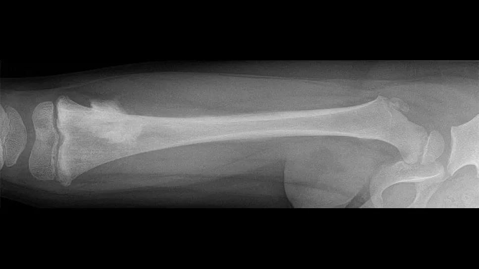
Image content: This image is available to view online.
View image online (https://assets.clevelandclinic.org/transform/8b25af4a-ae25-4ed9-a3ea-fc6cafefa620/xray-sarcoma-patient)
X-ray showing leg bones
Forty years ago, amputation was standard for young patients diagnosed with sarcoma in an arm or leg. Thanks to advances in imaging and chemotherapy delivery, limb salvage has become today’s conventional treatment.
Advertisement
Cleveland Clinic is a non-profit academic medical center. Advertising on our site helps support our mission. We do not endorse non-Cleveland Clinic products or services. Policy
Now up to 90% of pediatric patients with sarcoma are candidates for limb-salvage surgery, says Zachary D. Burke, MD, an orthopaedic oncologist at Cleveland Clinic who specializes in pediatric and adult sarcoma.
“There are some unique aspects to limb-salvage cases in pediatric patients,” he says. “First, kids are still growing. So, how do we account for future growth in a limb, particularly when a growth plate needs to be removed? How do we manage differences in limb length? Second, how do we ensure durability of a limb implant or reconstruction, allowing the patient to retain a high level of activity for 10, 20, 30 or more years down the road?”
Dr. Burke discussed these challenges and the latest advancements in addressing them in an episode of Cleveland Clinic’s Cancer Advances podcast.
Pediatric limb salvage is not a one-size-fits-all approach, says Dr. Burke. Every case requires a creative solution to a unique and sometimes difficult problem.
“Historically, reconstructing a limb that has had a big segment of bone and muscle and other tissue taken out involved implanting either a large metal prosthesis, called an endoprosthesis, or using a large segment of donor bone, called allograft,” he says. “Allografts can include bone, joint surface, tendons and soft tissue attachments. Endoprostheses are more commonly used now, but allografts may still be used in some cases.”
More recently, however, so-called biologic techniques have drawn more attention.
“There’s nothing as durable as native bone,” says Dr. Burke. “Reconstructions in which a child’s bone heals into viable, living bone provides the strongest reconstruction and durability.”
Advertisement
One biologic technique involves using a free vascularized fibular graft. In collaboration with a specialized plastic surgery team, a piece of the patient’s fibula along with its blood vessels is carefully removed. This graft is implanted into the bone defect in the leg or arm, and the blood vessels are reconnected.
“Because it uses a viable, vascularized piece of bone, this type of reconstruction is very durable,” says Dr. Burke. “There’s also a version of this technique that involves epiphyseal transfer, where we remove the fibula with its top growth plate. This allows the bone graft to continue growing after transplant.”
This can be combined with donor bone segments to perform complex reconstructions that save patients’ native joints.
A newer biologic technique is bone transport. This option draws on bone’s regenerative potential, which is particularly robust in children. Following surgical fixation and long-term immobilization and guided growth, a segment of bone can heal over time, providing a naturally durable reconstruction.
None of these solutions is perfect, however, cautions Dr. Burke.
“When you take a big piece of bone out of a kid’s leg, they do have repercussions and potentially complications,” he says. “When talking with a patient and their family about limb salvage, we do need to tell them what to expect from different techniques, the upside and the downside.”
Returning to the operating room is sometimes necessary, even decades after a patient’s reconstruction. Infection is one of the “dreaded complications,” he notes.
Advertisement
“When a prosthesis or piece of donor bone gets infected, it is a significant challenge to treat it without taking it out,” says Dr. Burke. “That becomes another limb-threatening situation. It’s frustrating for a patient to get through cancer and limb-salvage surgery only to get an infection years later that results in amputation.”
Dr. Burke’s work involves not only caring for sarcoma patients, but also leading a research lab focused on improving sarcoma outcomes and better treating complications like infections — whether with conventional antibiotics or newer molecular and immune therapies.
Another concern is implant failure, which can include fracture of the bone implant or loosening of the graft. This complication requires revision surgery. The more revisions required, the higher risk of infection and loss of function.
“Biologic fixation, where bone heals directly to an implant, can help prevent aseptic loosening,” says Dr. Burke. “Another durable option, particularly in patients who are a little older, is bone growth into a metal implant. Compressive osseointegration uses the bone’s physical property to load across the bone and induce growth into an implant. Another technique press fits the implant tightly into the bone to allow bone to grow onto and into the metal.”
Another important advancement in pediatric limb salvage is growing implants. Early iterations of growing implants involved a manual telescope mechanism. As the patient grew, the surgeon would lengthen the prosthesis during a surgical procedure, either adding a new piece onto the prosthesis or ratcheting and extending the implant.
Advertisement
Today’s noninvasive growing implants contain a magnetic coil. The length of the implant is extended simply by applying a magnetic field. Lengthening occurs slowly, millimeters at a time, and helps minimize leg-length differences.
Other advancements improving limb-sparing treatment for pediatric sarcoma are image guidance and 3D printing. CT and MRI scans are being used to produce both patient-specific implants and instrumentation.
“This has really come into play in orthopaedic oncology in the last five to 10 years,” says Dr. Burke. “We now can create custom cutting jigs that fit precisely onto a patient’s anatomy. We then can cut out a tumor with a degree of precision that we didn’t have before, minimizing the morbidity of resection and being more precise with our margins, and then inserting an implant or graft designed specifically for the patient.”
Emerging imaging technology may help surgeons not only navigate instruments during surgery, but also identify tumor cells, tag nerves and otherwise offer guidance for a more precise resection.
“We’ve come a long way in the last 40 years. All of these advancements are helping improve long-term outcomes for patients with sarcoma,” says Dr. Burke. “I’m excited to help advance care for these patients and see where this field goes in the next 30 to 40 years.”
Advertisement
Advertisement

The rare and aggressive form of bone cancer requires specialized treatment, but new and emerging therapies are providing hope for patients

Study also finds that 26% of children with cancer have mutations in DNA repair genes

Research highlights promising outcomes for treating recurrent and metastatic cases

Case study of radial-to-axillary nerve transfer for tumor-related deltoid nerve injury

Personalized reconstruction is an alternative to leg amputation or flail limb

Complex disease requires a comprehensive approach

Soft tissue pathologist discusses research into incorporating genomic data to improve risk stratification

Rare genetic disorder prevents bone mineralization