Locations:

A century-old medical tradition inspired the weekly complex-case reviews in Rheumatology
By Adam Brown, MD, and Taylor Koenig, MD
Advertisement
Cleveland Clinic is a non-profit academic medical center. Advertising on our site helps support our mission. We do not endorse non-Cleveland Clinic products or services. Policy
In our digitally focused world, user experience is important, but it isn’t limited to computers. At Cleveland Clinic’s Department of Rheumatic and Immunologic Diseases, medical fellows employ a century-old tradition – interactive case teaching – to hone medical trainees’ clinical reasoning and deepen their diagnostic skills.
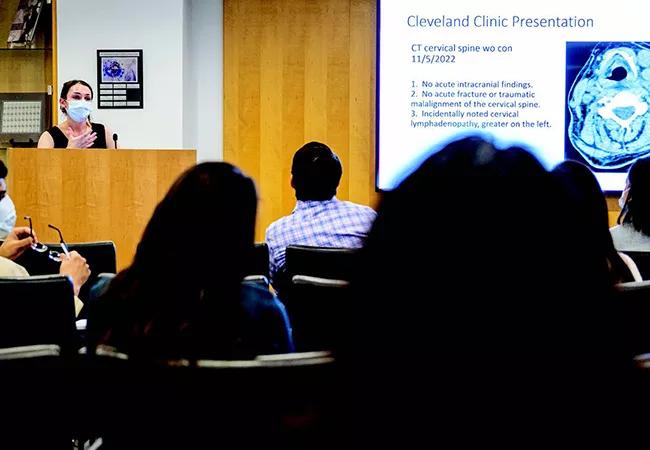
Each Friday at the R.J. Fasenmyer Center for Clinical Immunology, residents, fellows and faculty gather for Case Conference, the presentation of a clinical case with sufficient complexity to ignite educationally relevant debate and discussion.
The clinicopathologic conference was introduced at Harvard Medical School around 1900, spurred by student Walter B. Cannon’s interest in replacing some of the hours spent listening to lectures with purposeful case conversations. Richard Cabot, MD, then began using the method with third-year medical students at Massachusetts General Hospital.
In Rheumatology at Cleveland Clinic, fellows rotate the responsibility of presenter, choosing a case and establishing the facts: the presenting illness and patient’s medical history, as well as details from the physical exam. The trainees and faculty then discuss and debate differential diagnosis, what next tests and imaging should be pursued, and what treatments may be implemented. The presenter eventually reveals the final diagnosis.
The process makes for a meaningful educational activity because students move step by step through the reasoning they will use every day in practice. When a patient comes to them, what will they do about it? How will they prove one diagnosis or disprove another? They also are typically learning about complex cases, and often those requiring a multidisciplinary approach. Physicians from a variety of specialties often are part of our case discussions.
Advertisement
Case Conference offers an especially rich experience in rheumatology, which can be a confounding and humbling specialty. Following is a case discussed in in our Friday conference.
We presented the case of a 36-year-old male with a history of hyperlipidemia and a childhood penicillin allergy who presented to the Rheumatology Clinic with polyarticular joint pain and bilateral eye redness.
This case required collaboration among specialty providers and highlights the multidisciplinary nature of rheumatic disease. The patient’s symptoms started with sore throat and general malaise fewer than two weeks before his initial rheumatology evaluation. His throat culture was positive for streptococcus. He improved on antibiotic treatment with doxycycline. However, 10 days after symptom onset, the patient developed severe polyarticular joint pain and stiffness along with bilateral eye redness.
The patient was started on twice-daily naproxen by his primary care physician (PCP). On day 13, his symptoms continued to progress, and his PCP referred him to an emergency department (ED) for further evaluation.
In the ED, laboratory studies, including complete blood count and comprehensive metabolic panel, were within normal limits, with the exception of elevated C-reactive protein (CRP) at 5.7 mg/dL (normal < 0.9 mg/dL). Electrocardiogram revealed no ST changes and a normal PR interval. Urine tests for gonorrhea and chlamydia were negative. The patient was discharged with referrals to rheumatology and ophthalmology.
Advertisement
The patient was evaluated in the Rheumatology Clinic 14 days after symptom onset. He endorsed pain and stiffness in his hands, wrists, elbows, shoulders, knees, ankles and feet, which progressed in an additive fashion. His pain and stiffness had improved since starting naproxen. He denied eye pain, photophobia and vision loss associated with his eye redness. The patient denied rash, urethral discharge, dysuria, chest pain and gastrointestinal symptoms.
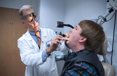
On exam, the patient had bilateral conjunctival erythema. He had synovitis and limited range of motion in his ankles, left wrist and bilateral third PIP joints. He also had tenderness with decreased range of motion in his hands and elbow without swelling. Dermatologic exam was pertinent for hyperpigmented patches over the patient’s anterior lower legs – findings that were confounded by a recent episode of poison ivy.
Because acute rheumatic fever was on the differential for this patient, we ordered an echocardiogram to evaluate for subclinical carditis and referred the patient to Infectious Disease as well as our ophthalmology colleagues, where he was diagnosed with episcleritis. Echocardiogram revealed normal systolic function, no valvular disease, and no pericardial effusion, ruling out subclinical carditis.
For our patient presenting with symmetric polyarthritis and episcleritis in the setting of a recent streptococcal infection, the two main diagnoses debated were acute rheumatic fever (ARF) and poststreptococcal reactive arthritis (PSRA). During the conference, we discussed how the diagnosis of ARF is distinguished from PSRA and how it may change management. We discussed the Jones diagnostic criteria for ARF; the patient only met one major (polyarthritis) and one minor (elevated CRP), making AFR less likely.
Advertisement
Additionally, the classic inflammatory arthritis of ARF is migratory: One joint is involved for a few days, resolves, then a different joint becomes involved. The joint pattern typically seen in PSRA is an additive, symmetric pattern like this patient described. Importantly, the joint pain in ARF also typically doesn’t start until two to three weeks after the sore throat. In contrast, PSRA often starts within two weeks of the sore throat, similar to our patient. The episcleritis is more non-specific and can be seen in both conditions, so this doesn’t narrow the diagnosis.
Distinguishing ARF and PSRA is important, because there are treatment implications. In ARF, we would expect the joint pain to be self-limiting, but recurrence of streptococcal infection would put the patient at risk for valvular heart disease, and prophylactic antibiotics may be initiated. In contrast, in PSRA we would not expect cardiac complications, but the articular manifestations may become more chronic, requiring immunosuppressive therapy.
With this multidisciplinary evaluation, our patient was diagnosed with PSRA. He has improved on naproxen and will follow up in the Rheumatology Clinic for further management.
This case demonstrates the interdisciplinary nature of rheumatic disease. Clinicians in primary care, infectious disease, ophthalmology and rheumatology all were essential in making this diagnosis and initiating the correct treatment for our patient.
Advertisement
Advertisement

Maintaining connections leads to referrals, recruitment and more
A commitment to sharing expertise has fostered a global exchange of ideas

Continuous Improvement model makes success repeatable

Medical students will complete five weeks of clinical training

Findings about the tool's utility were presented at the American Society of Nephology’s 2022 Annual Meeting

The program provides an inside look and hands-on experience for high school students interested in nursing

Join us in New York City Dec. 5-6

Program helps participants experience nursing through hands-on exploration