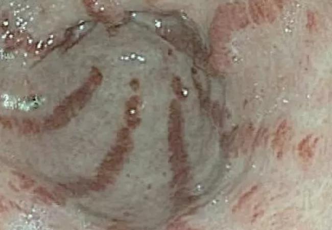
Gastric antral vascular ectasia in systemic sclerosis and serositis in antisynthetase syndrome

By Soumya Chatterjee, MD, MS, FRCP
Advertisement
Cleveland Clinic is a non-profit academic medical center. Advertising on our site helps support our mission. We do not endorse non-Cleveland Clinic products or services. Policy
At the 2018 American College of Rheumatology Annual Meeting in Chicago, Cleveland Clinic rheumatology fellows presented research on two rare autoimmune conditions. Their findings shed new light on these little-understood diseases.
Second-year rheumatology fellow Rabeea Mirza, MD, compared gastric antral vascular ectasia (GAVE) in systemic sclerosis (SSc) with GAVE in other diseases.
Background. GAVE is a pathologic angioectasia with a characteristic endoscopic appearance. Rugal folds with dilated blood vessels radiate from the antrum and converge at the pylorus, resembling watermelon stripes, supporting the name “watermelon stomach.” GAVE can cause anemia and significant morbidity, hence there is need for surveillance.
GAVE has been associated with cirrhosis of liver, autoimmune diseases (e.g., SSc, rheumatoid arthritis, primary biliary cholangitis), end-stage renal disease, hypertension, heart failure, hypothyroidism and chronic pulmonary disease. It also can occur after hematopoietic stem cell transplantation. Prevalence of SSc-associated GAVE is highly variable, ranging from 1 to 76 percent of patients with SSc. Prevalence of GAVE in other associated diseases and its long-term outcomes are still unknown.
Methods. We conducted a retrospective chart review of patients with GAVE and evaluated those diagnosed between 2012 and 2017. We initially identified 145 GAVE patients and separated them into cohorts of those with SSc and those with other diseases. We selected 37 consecutive SSc and 37 consecutive non-SSc patients from the GAVE database. Outcomes were defined by number of transfusions, number of recurrences of GAVE bleeding diagnosed endoscopically and death.
Advertisement
Results. This study demonstrated that SSc patients with GAVE were significantly younger than those with non-SSc GAVE, and were mostly females. Patients were followed for a median of five years.
Clinical manifestations associated with GAVE in both SSc and non-SSc groups were telangiectasias, melena, hematemesis, fatigue, dyspnea and lightheadedness. When adjusted for pre-transfusion hemoglobin, difference in transfusion requirements was not statistically significant between the two groups. There was no difference in use of NSAIDs and anticoagulants between the two groups. There also was no difference in number of recurrences of GAVE. Two patients with cirrhosis of liver died.
Further studies with larger cohorts of GAVE patients may be helpful in understanding its natural history and outcomes in specific diseases.
First-year rheumatology fellow Alexis Katz, DO, studied the prevalence of serositis in antisynthetase syndrome (ASS), its clinical significance and its association with specific ASS autoantibody (Ab) subtypes.
Background. ASS is a relatively rare autoimmune disease characterized by interstitial lung disease, myositis, inflammatory arthritis, Raynaud phenomenon and mechanic’s hands. Eight autoantibodies to aminoacyl-transfer RNA synthetases have been described so far: Jo-1, PL-7, PL-12, EJ, OJ, YRS, KS and Zo. Morbidity and mortality in ASS is mainly related to pulmonary complications. However, little has been reported about the prevalence of serositis (pleural and/or pericardial effusions) in ASS other than in small cohort studies (15-20 patients) and case reports.
Advertisement
Methods. Clinical data were obtained by retrospective review of electronic medical records from 2004 to 2017. Our study included patients diagnosed with ASS by a rheumatologist. All patients had one of the following ASS antibodies: Jo-1, PL-7, PL-12, EJ or OJ. Pleural effusions were qualified as trace, small, medium or large, based on chest radiographs and thoracic CT scans. Pericardial effusions were classified as trace, small, medium, large or tamponade, based on echocardiographic findings.
Results. A total of 93 patients were included in this study. The mean age was 57.5 years; 63 percent were females.
Out of 90 patients with complete data available, 42.2 percent had pleural effusion(s) and 47 percent had a pericardial effusion, of which 10 percent were moderate to large. One patient had tamponade physiology. Anti-Jo-1 patients were significantly less likely to have pleural effusions when compared to patients with other antibodies. Anti-PL-12 had a higher frequency of pleural effusions relative to anti-Jo-1, anti-PL-7 and all other antibodies combined.
More research is necessary to better understand, diagnose and treat both GAVE in SSc and serositis in ASS. Our work is intended to raise awareness of these conditions, share new insights and serve as a springboard for further investigation.
Dr. Chatterjee directs the Scleroderma Program in the Department of Rheumatic and Immunologic Diseases.
Advertisement
Advertisement

The case for continued vigilance, counseling and antivirals

High fevers, diffuse rashes pointed to an unexpected diagnosis

No-cost learning and CME credit are part of this webcast series

Summit broadens understanding of new therapies and disease management

Program empowers users with PsA to take charge of their mental well being

Nitric oxide plays a key role in vascular physiology

CAR T-cell therapy may offer reason for optimism that those with SLE can experience improvement in quality of life.

Unraveling the TNFA receptor 2/dendritic cell axis