Locations:





Innovative procedure offers less-invasive alternative to Latarjet procedure
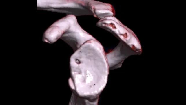
Image content: This image is available to view online.
View image online (https://assets.clevelandclinic.org/transform/c4adafd7-4b91-461e-86f3-d05059348afb/glenoid-bone-loss)
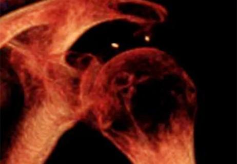
CT image demonstrating glenoid bone loss
Shown above: CT image demonstrating glenoid bone loss
Advertisement
Cleveland Clinic is a non-profit academic medical center. Advertising on our site helps support our mission. We do not endorse non-Cleveland Clinic products or services. Policy
For patients with recurrent anterior shoulder instability, repeated dislocations can do more than damage the labrum. Over time, they can cause clinically significant glenoid bone loss, leaving too little bone for a soft-tissue repair alone to be effective.
In that setting, orthopaedic surgeons have traditionally turned to the Latarjet procedure. But arthroscopic bone augmentation is emerging as a less-invasive option for selected patients, with the potential to restore glenoid bone stock while also addressing associated intra-articular pathology.
“When I am concerned about instability with possible glenoid bone loss, I typically order a CT scan with 3D reconstruction,” says Cleveland Clinic orthopaedic surgeon Joseph Tramer, MD. “That gives me a fairly accurate measure of the percentage of bone loss.”
This is a standard instability workup when there is concern for bone loss, he notes, because isolated soft-tissue repair has poor outcomes when bone loss is significant. Identifying and quantifying bone loss preoperatively can help avoid failed stabilization.
The Latarjet procedure has a long track record and is highly effective at preventing recurrent instability. Due to its decades of research data, it remains the gold standard in some populations, especially young athletes participating in contact sports.
However, it is an open, technically demanding procedure. It involves transfer of the coracoid to the anterior glenoid, typically through a split in the subscapularis, with screw fixation.
Advertisement
“It works well to treat shoulder instability, but it’s a fairly traumatic operation to the muscle compared with an arthroscopic approach,” Dr. Tramer says.
Risks include hardware irritation, screw breakage and complexity due to nearby neurovascular structures. These limitations have helped drive interest in arthroscopic bone block procedures.
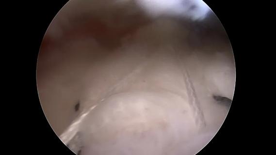
Arthroscopic bone augmentation, or arthroscopic glenoid reconstruction, is not new but is gaining popularity, building on techniques established in labral repair. It is performed through small portals with a camera.
“Instead of open surgery, where you can only see a portion of the shoulder, I put the camera in and can see the entire shoulder,” Dr. Tramer says.
The procedure preserves the subscapularis by passing the graft around it rather than through it. Instead of transferring the coracoid, it uses an allograft or autograft bone block to reconstruct the anterior glenoid.
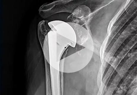
The graft Dr. Tramer uses is an off-the-shelf, preshaped, predrilled allograft implant, which simplifies graft preparation. Fixation is achieved with smaller suture, button-based constructs rather than traditional screws.

Image content: This image is available to view online.
View image online (https://assets.clevelandclinic.org/transform/e94af746-43e2-41b7-b481-51892efba75f/glenoid-reconstruction)
X-ray after arthroscopic glenoid reconstruction, showing button-based fixation.
The goal is for the graft to heal to the native glenoid and incorporate over time. Patients typically remain in a sling for about one month. Range-of-motion work typically begins during months 2 and 3. Lifting is generally restricted until about month 3, with return to sports and contact activity around month 6.
Arthroscopic visualization also makes it easier to address other pathology that commonly accompanies recurrent instability, including labral injury, capsular pathology and Hill-Sachs lesions.
Advertisement
“Arthroscopy allows me to inspect and treat all of these in one setting,” Dr. Tramer says. “It’s much easier to repair the labrum over the bone block and perform remplissage for a Hill-Sachs lesion at the same time. Doing that alongside an open Latarjet can require combined open and arthroscopic approaches.”
In Dr. Tramer’s practice, arthroscopic bone augmentation is best suited to selected patients with:
Latarjet remains preferred in some high-risk groups because it has the strongest long-term outcomes data. Although arthroscopic bone augmentation is promising in those groups as well, the long-term evidence is not yet as robust. Broader adoption will depend on continued outcomes research and follow-up, Dr. Tramer notes.
Still, for patients with recurrent instability, “it helps to talk to someone with as many tools in the toolbox as possible,” he says. “With growing clinical experience and expanding outcomes data, arthroscopic bone augmentation is poised to play an increasingly important role in the management of recurrent shoulder instability.”
Advertisement
Advertisement
Why shifting away from delayed repairs in high-risk athletes could prevent long-term instability and improve outcomes
Conventional arthroplasty performs better than reverse arthroplasty for patients with an intact rotator cuff
Sinus tracts can occur years later and not near the incision site
CT imaging using radiopaque markers can help assess healing after surgery
Iliac crest structural autograft repairs large, uncontained defect
Ultrasound-guided debridement for selected patients with persistent symptoms
Cleveland Clinic’s new program uses a cross-institute model to provide complete care for women of all ages and athletic levels
A practical look at patient selection, implant choice and the unanswered questions around long-term value
 Search