Locations:

Is it lupus?
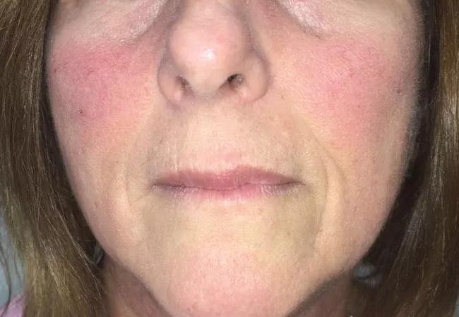
Image content: This image is available to view online.
View image online (https://assets.clevelandclinic.org/transform/6331d244-da4f-4d79-958d-d91964c3fc72/20-RHE-1935445-Chatterjee_Parvovirus-associatedArthritis-CQD-650x450-1_jpg)
20-RHE-1935445-Chatterjee_Parvovirus-associatedArthritis-CQD-650×450
By Soumya Chatterjee, MD, MS, FRCP
Advertisement
Cleveland Clinic is a non-profit academic medical center. Advertising on our site helps support our mission. We do not endorse non-Cleveland Clinic products or services. Policy
A 58-year old female with a history of cervical spondylosis and hypothyroidism, presented with a two-week history of joint pain.1 She reported that her pain and swelling began in the right knee, migrated to the right ankle, and finally to the metacarpophalangeal (MCP) and proximal interphalangeal (PIP) joints of her fingers.
On exam, the patient was afebrile, with a malar rash (figure). She had swollen and tender MCP and PIP joints. She had difficulty making a fist and extending her fingers.
Approximately two weeks before the patient’s symptoms began, her 5-year-old grandson developed a febrile illness with headache, body aches, malaise, and rash on the cheeks, trunk, and extremities, but no joint symptoms. The patient’s 10-month old granddaughter developed a similar illness ten days later. Eight days before the patient presented, her 25-year-old daughter developed headache and malaise, with pain and stiffness of the wrists and knees, but no rash or fever. All the symptoms of her family members resolved spontaneously within seven days.
Initial laboratory studies revealed a mild normochromic normocytic anemia with a hemoglobin level of 11.5g/dL, normal white blood and platelet counts, and normal results for a comprehensive metabolic panel. She had a positive antinuclear antibody (ANA) result (1:160 [homogeneous pattern; negative <1:80 serum dilution]). Her rheumatoid factor was negative; levels of inflammatory markers (ESR and C-reactive protein) were normal.
It was felt that with the pertinent recent family history, it would be premature to give her a diagnosis of a chronic autoimmune rheumatologic disease, such as systemic lupus erythematosus (SLE) or rheumatoid arthritis (RA). On the other hand, the recent family history of exposure to children with fever and a facial rash suggested a possible diagnosis of erythema infectiosum (fifth disease).
Advertisement
Further laboratory studies revealed significant titers of IgM (15.04 index value) and IgG (6.45 index value) antibodies to parvovirus B19. Antibodies to cyclic citrullinated peptide (anti-CCP), double-stranded DNA, and extractable nuclear antibodies were all negative. The patient’s symptoms resolved with prednisone 35 mg daily, which was tapered over a week. Six months later, repeat laboratory studies were negative for IgM antibodies to parvovirus B19 and ANA.
Erythema infectiosum is common in children and is caused by human parvovirus B19. The infectious phase of the virus generally begins 24–48 hours before the earliest detectable symptoms and lasts until the associated rash resolves. Symptoms include fever, headache, sore throat, itching, cough, upset stomach, sneezing, conjunctivitis, and muscle aches. These flu-like symptoms last approximately 5–7 days before the classic “slapped cheek rash” develops (which was thought to be a “malar rash” in our patient), which is sometimes followed by a maculopapular rash on the rest of the body. Although rare in children, joint symptoms are common in adults and tend to occur more frequently in women than men.2 The non-erosive, often symmetric, arthropathy generally last for 1–3 weeks;2 however, they can be more protracted and may mimic the onset of a systemic autoimmune rheumatologic disease such as RA or SLE. Both ANA and rheumatoid factor can be transiently positive, further confusing the picture. Other viral infections that can cause arthralgia and arthritis include Rubella, Hepatitis B and C, HIV, Chikungunya, Dengue, Ebola, Adenovirus, Enteroviruses and Herpesviruses.
Advertisement
Note: Image used with permission. Originally published in: Chatterjee S. Malar rash and polyarthritis. JAMA. 2019;321(3):303-304.
Advertisement
Advertisement

Going beyond simple referrals to streamline patient access

Tracing our understanding of this complex systemover more than a century

Drug-free remission holds more than a year after treatment

Cleveland Clinic group works toward earlier diagnosis of this rare disease

Researching the biological basis for why treatment is or is not effective

Scribing system helps create more face-to-face interactions

A conversation with Leonard Calabrese, DO

The case for continued vigilance, counseling and antivirals